apolysis Separation of old cuticle from underlying epidermal cells caused by release of ecdysteroids at the beginning of each molt.
ametabolous Characterized by no significant metamorphosis. Immature body form does not differ from adult, except for external genitalia and number of stages is variable.
ecdysis The act of shedding the old cuticle.
hemimetabolous Immature forms closely resemble adult form and pass through a fixed number of juvenile instars; last instar exhibits wing buds and incomplete metamorphosis.
holometabolous Immature forms are vermiform, with a fixed number of larval instars; complete metamorphosis during pupal stage.
molt Period during which the insect synthesizes new cuticle and other structures appropriate for next developmental stage; begins with ecdysteroid-induced apolysis and ends with ecdysis of old cuticle.
intermolt Period of feeding and growth; begins with ecdysis from previous stage and ends with apolysis.imaginal discs Clusters of undifferentiated embryonic cells in holometabolous insects that proliferate during larval stages, then differentiate during the pupal stage upon induction by ecdysteroids in the absence of juvenile hormone.
USP Nuclear transcription factor related to the retinoid-X-receptors; dimerizes with the ecdysteroid receptor once it is liganded and binds as a complex to DNA to regulate gene expression.
INTRODUCTION
The majority of insects undergo embryonic development within an egg, then advance through a series of immature larval stages culminating in metamorphosis to the adult, reproductive form. It has been suggested by Lynn Riddiford and James Truman that the stunning evolutionary success of insects is attributable to complete metamorphosis, whereby the immature larva, essentially a gut covered with cuticle, is exquisitely adapted for resource exploitation, rapid growth, and avoidance of competition with its conspecific adult, reproductive stage. Metamorphosis is a magnificent transformation of one body form into a completely different one under the control of hormones. Upon reaching the requisite body size, hormonal signals are released, committing the animal to a postembryonic rebirth. Carroll Williams summarized this basic developmental process with the following anecdote:
“The earth-bound stages built enormous digestive tracts and hauled them around on caterpillar treads. Later in the life-history these assets could be liquidated and reinvested in the construction of an entirely new organism—a flying machine devoted to sex.”
Conversion of the energy accumulated by the larval form into the adult during metamorphosis is a fascinating process of organismal re-modeling. It is accomplished via programmed cell death of larva-specific cells, re-programming of others, and the postembryonic birth of new cells from imaginal disc tissues upon receipt of precisely timed and coordinated hormonal signals.
INSECT BODY PLANS AND DEVELOPMENTAL PROGRAMS
Three distinct patterns of growth are observed in insects, distinguished by body form and type of metamorphosis: ametabolous, hemi-metabolous, and holometabolous. Insect orders exhibiting ametabolous development belong to the Apterygota, or wingless insects. Included in this group are the primitive orders Protura, Collembola, and Diplura, whose earliest stages are miniature adults in form, except for the absence of external genitalia. They grow continuously and lack metamorphosis. Attainment of reproductive competence occurs at an indefinite time in development, and molting continues even after the adult stage is reached. Little is known about the hormonal control of development in the Ametabola.
The majority of insects are winged and have either partial or complete metamorphosis. The Hemimetabola such as grasshoppers and crickets emerge from the egg formed as small, immature versions of the adult, and are called nymphs. They lack wings and functional reproductive organs. After a series of molts, the number usually constant from one generation to another, nymphs pass directly to the winged, reproductive adult stage in a single step. This mode of development is referred to as incomplete metamorphosis. The more advanced insect orders including moths, beetles, flies, and wasps develop as vermiform (wormlike) larvae during the immature stages. The complete metamorphosis of these groups is a two-step process in which a sessile, non-feeding pupal stage is intermediate between larva and adult. The pupal stage allows for a complete change in body form from larva to winged, hexapod adult. This total transformation of body form during complete metamorphosis requires a high degree of postembryonic cellular programming under the control of hormones. Three types of cellular processes are dictated, mostly by hormonal signaling involving ecdysteroids and juvenile hormones (JHs). First, many larva-specific structures such as body wall muscles and neurons must be eliminated through a programmed cell death known as apoptosis. Second, some cells persist to the adult stage, but are extensively re-modeled to serve adult functions. Finally, new cells derived from imaginal discs are born.
The diversity of insect groups places limits on generalizations about hormonal control of development. The effects of hormones in one group may not be the same for other groups, because of different patterns of growth and cellular specification. In the Lepidoptera, for example, larval epidermal cells change their cuticle secretion program at metamorphosis and switch to production of pupal and subsequently adult cuticle. Exogenous JH application at this time maintains the larval secretory program, resulting in supernumerary (extra) instars. Similarly, properly timed application of JH in the early pupal stage of moths can cause a second pupal stage. However, the fruit fly Drosophila and other higher Diptera are largely resistant to such effects of JH. This may relate to differences in the developmental program of fly epidermal cells. The entire epidermis in the head and thorax is programmed for secretion of larval cuticle only, and dies at metamorphosis. It is replaced by imaginal disc tissue, which remains undifferentiated throughout larval life in the presence of JH. Abdominal epidermal cells also die after pupation and are replaced by abdominal histoblasts. Thus, the development program of epidermis in higher flies is entirely distinct from the Lepidoptera, and its lack of response to JH in the early stages may be of consequence of this very different design.
These differences in responses to hormones complicate interpretation of many findings, especially because it has become fashionable to test overall hypotheses using the moth Manduca for physiology and endocrinology experiments, and Drosophila for genetic manipulations. Generalizing about common mechanisms for these two evo-lutionarily distant groups should be done cautiously.
INSECT DEVELOPMENTAL
HORMONES—DEFINITIONS AND
CLASSIFICATION
Ecdysteroids are relatively polar steroid hormones released by the prothoracic glands in immature stages and the gonads in adults. They are the chief regulators of gene expression in insect development and reproduction. The first structure to be elucidated was alpha-ecdysone (aE), the immediate precursor to 20-hydroxyecdysone
(20HE), accepted as the main protagonist in ecdysteroid actions. While 20HE indeed is associated with the vast majority of ecdyster-oid actions, there is evidence to support aE as a signaling molecule in certain instances. Other suspected ecdysteroids are 20,26HE and makisterones.
Juvenile hormones (JH), sesquiterpenoid derivatives from the sterol synthesis pathway, are released by the corpora allata. Six types are known in insects, with JH-III being the predominant form. The chief action of JHs is to modulate ecdysteroid-mediated gene expression. No receptors for JH have been clearly identified, but it seems likely that the hormone interacts with intracellular receptors or proteins that modulate ecdysteroid signaling. The signature of JH action is to promote expression of the immature phenotype.
Prothoracicotropic hormone (PTTH) is a large peptide hormone released by brain neurosecretory cells from terminals in the corpora cardiaca or corpora allata. PTTH stimulates the prothoracic gland to synthesize and release aE or in some instances 3-dehydroecdysone, which then are converted to 20HE in the hemolymph or by target tissues. PTTH is a homodimer consisting of two 12 kDa subunits joined by a disulfide bond. Release of PTTH is regulated by sensory inputs to the brain, which convey information about body size and nutritional state.
Bursicon is a 30-40 kDa peptide that accelerates sclerotization of cuticle. It is released from neurosecretory cells in the brain and ventral nerve cord after each ecdysis. Since insects are particularly vulnerable to predation during and after ecdysis, rapid hardening of the cuticle maximizes survival.
Eclosion hormone (EH) is a 62-amino acid peptide that mediates circadian-mediated eclosion to the adult stage and each larval ecdysis. EH is released into the blood from ventral median neurosecretory cells of the brain, causing release of ecdysis-triggering hormone (ETH) from Inka cells of the epitracheal endocrine system. It is also implicated in the elevation of cyclic GMP in a subset of neurons in the central nervous system (CNS) which initiates ecdysis behavior.
Ecdysis-triggering hormones are peptides synthesized and released from Inka cells at the end of the molt. ETHs act directly on the CNS to cause a behavioral sequence that leads to shedding of the cuticle. ETH release is caused by EH, and the action of ETH on the CNS leads to release of EH from VM neurons. It has been proposed that ETH and EH engage in a positive feedback signaling pathway that results in depletion of ETH from Inka cells, which is necessary for the transition from pre-ecdysis to ecdysis behaviors.
MODES OF HORMONE ACTION
Hormones are chemical messengers that travel throughout the body to effect responses in specific tissues. Targeted cells have receptors that, upon binding the hormone, transduce the signal into a cellular response. Insect hormones regulate development either by activation of intracellular receptors or receptors at the cell membrane.
Ecdysteroids and JHs Activate Intracellular Receptors
Ecdysteroids and JHs are relatively lipophilic signaling molecules able to easily traverse the cell membrane. Upon entry, they bind to intracellular proteins called nuclear receptors or nuclear transcription factors, which reside either in the cytoplasm or in the nucleus. Regardless of their initial location, hormone binding triggers passage to the nucleus, where the receptor forms a complex with other proteins and then binds directly to DNA, inducing or repressing gene expression.
Several factors govern diverse, stage-specific responses of target cells to ecdysteroids. First, ecdysteroid receptors occur as multiple subtypes, including EcR-A, EcR-B1, and EcR-B2. The response to ecdysteroids is governed by the subtype expressed by target cells, as well as which subtype of USP (USP-1 or USP-2), the EcR partner, is expressed. The transient availability of receptors in target cells leads to sensitive periods at specific stages of development. For example, both EcR-A and EcR-B1 are present throughout larval life, but the ratio of the two favors EcR-B1. In contrast, levels of EcR-A increase during metamorphosis. Second, the affinity of EcR subtypes can vary, as can the kinetics of a given response to an ecdysteroid peak. These patterns of receptor and dimer partner expression appear to mediate different cellular responses to ecdysteroids at different developmental times.
Peptide Hormones Activate Cell-Surface Receptors
Most peptide hormones bind to a class of integral membrane proteins on the plasma membrane of the target cell. The majority of these are G-protein-coupled receptors (GPCRs) that trigger intrac-ellular second messenger cascades.
EMBRYOGENESIS
Distinct patterns of embryonic development are observed in hemimetabolous and holometabolous insects. In both cases, the embryo produces multiple cuticular layers, and the appearance of these coincides with pulses of ecdysteroids. JH levels are generally low during early embryogenesis, but climb later to program nymphal or larval cuticle formation upon appearance of an ecdysteroid peak.
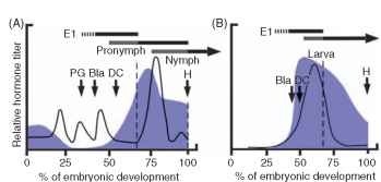
In the hemimetabolous grasshopper Locusta, four peaks of ecdys-teroids are observed, corresponding to production of serosal cuticle and three embryonic cuticle layers (Fig. 1A). JH levels are high immediately after oviposition, because of maternal contribution to the yolk, but rapidly decreases to low levels. At ~20% of embryonic development and before prothoracic glands are developed, the first ecdysteroid peak consisting exclusively of aE occurs in the presence of relatively low JH levels. This first peak comes from the release of maternal ecdysteroids stored as polar conjugates, and shortly thereafter the serosal cuticle is secreted. A second aE peak occurs at ~30% development, leading to formation of the first embryonic cuticle. A third ecdysteroid peak occurs just after differentiation of the protho-racic glands, and this coincides with the first appearance of 20HE and 20,26HE. Nevertheless, levels of aE together with 20,26HE predominate at this time, leading to the secretion of the second embryonic cuticle, which Truman and Riddiford refer to as pronymphal cuticle. It is the first layer of cuticle tough enough to require shedding via ecdysis behavior. The coincidence of the early, aE peaks with cuticle secretion suggest that aE is not only a 20HE precursor but also a biologically active hormone at certain times of development. At 70% development, the first embryonic cuticle is shed, followed quickly by a large peak of ecdysteroids. This is the first exposure of the embryo to substantial 20HE levels in the presence of JH, leading to synthesis of the first instar nymphal cuticle. This peak of ecdysteroids contains large amounts of aE, 20HE, and 20,26HE.
At hatching, the grasshopper emerges from the egg under the ground, still surrounded by the pronymphal cuticle. Despite its hexa-pod body plan, the animal exhibits a classic vermiform (wormlike)

FIGURE 1 Hormone levels during embryonic molts in a hemi-metabolous insect, the grasshopper Locusta migratoria (A) and in a holometabolous insect, the moth, Manduca sexta. During Locusta embryogenesis, four peaks of ecdysteroid are observed, each corresponding to secretion of a layer of cuticle. The first peak of predominantly aE at 20% development initiates secretion of the serosal cuticle (not shown). A second peak prior to prothoracic gland (PG) differentiation causes secretion of the first embryonic cuticle (E1). Just after blastokinesis (Bla), a third ecdysteroid peak leads to secretion of the pronymph cuticle. Shading of the pronymph time line at the top of the plot indicates the pharate stage, which ends with ecdysis of the E1 cuticle (vertical dotted line). The fourth ecdysteroid peak, occurring for the first time in the presence of JH, contains approximately equal amounts of aE, 20HE, and 20,26HE. This causes secretion of the first-stage nymphal cuticle. At hatching the nymph sheds the pro-nymph cuticle upon escaping from the substrate. Secretion of the first embryonic cuticle in Manduca occurs in the absence of an ecdyster-oid peak. The first larval cuticle is secreted in response to elevated ecdysteroids in the presence of JH just after dorsal closure (DC). Note that JH levels in Manduca rise earlier in development than in Locusta and that ecdysteroid signaling always occurs in the presence of JH.
locomotory pattern as it escapes the egg pod and maneuvers through the substrate to the surface. In a matter of seconds to minutes, the pronymphal cuticle is shed, the animal stretches its legs, and switches abruptly to hexapod behavior. Truman and Riddiford have called attention to many similarities between the hemimetabolous pronymph and the holometabolous larva, suggesting that the latter has resulted from a hormonal shift in embryogenesis, resulting in an extended postembry-onic phase of pronymph development. The ancestral pronymph undergoes an extended, multi-stage developmental sequence as a larva.
The importance of a JH-free period during early embryogenesis of hemimetabolous insects (grasshopper—Schistocerca, cricket— Acheta) has been demonstrated by treatment of eggs with JH analogs. This results in inhibition of blastokinesis, reduction in the number of embryonic cuticle layers produced, premature appearance of nymphal cuticle and mouthparts, and reduced body size.
Embryogenesis in holometabolous Lepidoptera is somewhat simpler, with the secretion of only three cuticles, one serosal and two embryonic cuticles. Levels of both ecdysteroids and JH are unde-tectable early in embryogenesis, but rise earlier as compared to the Hemimetabola, or at about 30% development (Fig. 1B), preceding the ecdysteroid peak. Therefore, unlike hemimetabolous embryo-genesis, the first exposure to ecdysteroids occurs in the presence of JH, leading to production of the first larval cuticle. An embryonic ecdysis occurs at 70% development, and first instar larva hatching does not involve cuticle shedding. The importance of a JH-free period observed for Hemimetabolous insects is not the case for embryogenesis in the Holometabola. Embryos are largely insensitive to exogenous JH treatment, suggesting perhaps the absence of receptors for these hormones until later in embryogenesis.
LARVAL DEVELOPMENT: FEEDING, MOLTING, AND ECDYSIS
The Intermolt Because the exoskeleton places limits on growth, insect development occurs in stages, each ending with molting and cuticle shedding, or ecdysis. During the intermolt, which follows ecdysis, JH levels are maintained around 1-10|igml-1 in the blood (Fig. 2). It is presumed that these JH levels promote a high metabolic rate, active feeding behavior, synthesis of larval cuticle proteins, and continuous proliferation (but not differentiation) of imaginal discs. Considerable growth during the immature stages is possible because the immature integument is predominantly unsclerotized procuticle, which is quite flexible as compared to hard, sclerotized adult cuticle. Several mechanisms allow for larval cuticle expansion. The epidermal cells add new protein to the cuticle throughout the intermolt, increasing the

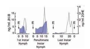
FIGURE 2 Hormonal regulation of development in postembry-onic stages of the cockroach Nauphoeta cinerea, representing hemimetabolous development. During the immature stages of Nauphoeta, JH levels are elevated each time ecdysteroid levels rise to initiate a molt, resulting in secretion of nymphal cuticle. Following ecdysis to the last instage.star nymph, JH levels drop precipitously. Adult commitment is signaled by a biphasic ecdysteroid peak in the absence of JH. Incomplete metamorphosis occurs during the period between ecdys-teroid elevation and ecdysis to the adult
surface area by intusseception. In addition, new cuticle in Manduca is deposited in vertical columns that are gradually re-oriented during the feeding stage to allow for expansion. The increase in size during the larval stage can be quite impressive, as in fifth instar Manduca, which increases its body weight from ~ 1 g on the first day of development to 15 g, and its cuticular surface area by approximately fivefold just prior to pupation. Blood-feeding insects such as Rhodnius are known to release serotonin after a blood meal, which acts as a plasticizing agent, facilitating the enormous expansion of the body wall after a blood meal.
At some point during each immature stage, growth results in a decision by the brain to initiate the molt. In Rhodnius, the simplest case known, stretch receptor input from abdomen to brain causes release of PTTH, which induces synthesis and secretion of ecdys-teroids from the prothoracic glands. In most insects, the decision to release PTTH is more complicated and less well understood, but it has to do with body weight, nutritional state, and time spent at that stage. The immediate effects of ecdysteroid elevation include cessation of feeding and apolysis, the detachment of the old cuticle from underlying epidermal cells. Apolysis of larvae results in head capsule slip, which occurs because the new head capsule is larger than the old one. This is the most visible sign that the molt has been initiated. If elevation of ecdysteroids occurs in the presence of JH, epidermal cells maintain the “status quo” secretory program for immature phe-notype, and larval cuticle is secreted (Fig. 2). Through the action of molting fluid, most components of the old cuticle are broken down and recycled into the new layer.
During the period of new cuticle synthesis, ecdysteroids also orchestrate gene expression crucial to the synthesis and action of peptide hormones that control ecdysis behaviors. Ecdysis is a complex process in which the old cuticle is shed not only from the surface of the animal but also from the lining of the foregut, hindgut, and the inner walls of the tracheal system. Success in this process depends on completion of new cuticular synthesis, attachment of the musculature to the newly deposited cuticle, and digestion of the old cuticle. In addition, the animal prepares for a sequence of Houdini-like escape behaviors necessary to shed the old cuticle. These consist of pre-ecdysis, ecdysis, and post-ecdysis behaviors. The ability to perform these behaviors depends on orchestration of a peptide-signaling cascade involving the CNS and the epitracheal endocrine system.
For the ecdysis-signaling cascade to be functional at the appropriate time, ecdysteroids orchestrate gene expression in four ways. First, genes are activated in epitracheal glands to increase production of ETHs in endocrine Inka cells. Second, the release of ETHs initiates ecdysis behaviors through direct action on the CNS. Although the CNS is not sensitive to ETHs during the feeding stage, acquisition of sensitivity occurs upon elevation of ecdysteroids, specifically around the time of apolysis. Third, the nervous system becomes competent to release EH, a peptide hormone that targets Inka cells to cause the release of ETH. Finally, elevated ecdysteroids exert a negative influence on the secretory competence of Inka cells. As long as ecdyster-oids remain high, Inka cells are unable to secrete ETHs in response to EH exposure. This latter effect of ecdysteroids, to block release of ETHs from Inka cells, may be designed to ensure that ecdysis does not occur prematurely. Declining ecdysteroid levels at the end of the molt provide the necessary signal permitting expression of genes needed for secretory competence.
It is believed that initiation of ecdysis behaviors occurs as a result of an ongoing conversation between the nervous system and Inka cells. In Manduca, the neuropeptide corazonin is released from the brain, causing the Inka cells to release small amounts of ETHs, initiating pre-ecdysis behavior. Pre-ecdysis behaviors are thought to loosen the remaining connections between the new and old cuticle. Circulating levels of ETH also target VM neurons in the brain to cause release of EH. EH acts back on Inka cells in a positive feedback manner to cause massive release of ETHs, triggering the transition from pre-ecdysis to ecdysis. It is thought that the action of ETH activates a downstream cascade of peptidergic signaling within the CNS to schedule each step of the behavioral sequence. Included in this cascade are neuropeptides called kinins and crustacean car-dioactive peptide (CCAP), the latter named for its initial discovery and biological activity. In the context of insect ecdysis, kinins appear to be the immediate chemical signals for pre-ecdysis initiation and
CCAP for activation of peristaltic ecdysis behavior. Upon escaping the old cuticle, the animal is surrounded by a new soft cuticle and is therefore extremely vulnerable to injury. In the case of newly eclosed adults, the wings must be expanded. The neuropeptide hormone bursicon plays dual roles in this process, inducing both cuticle extensibility necessary for wing expansion and subsequently sclerotization (hardening) of the new cuticle.
In summary, ETH, EH, kinins, CCAP, and bursicon regulate ecd-ysis at all stages. This includes embryonic ecdysis in Manduca, and adult eclosion.
METAMORPHOSIS: TRANSITION FROM IMMATURE TO ADULT
The transition from immature to adult is signaled by the elevation of ecdysteroid levels in the absence of JH. This is a one-step process in hemimetabolous insects. During the last nymphal instar of the cockroach Nauphoeta, JH levels fall from 5-10 |ig ml-1 to less than l|igml-1 prior to the next ecdysteroid peak (Fig. 2). Appearance of ecdysteroids at this low JH level signals a commitment to an adult gene expression pattern. Some examples of cellular responses to the adult commitment peak include mitosis in wing pad tissue, development of flight muscles, competence of gonadal accessory glands to differentiate, the formation of external genitalia, and re-organization of the nervous system to accommodate these new adult structures. Also included are more subtle alterations, such as the relative proportions of body parts and addition of secondary sexual characteristics such as acoustic organs for communication.
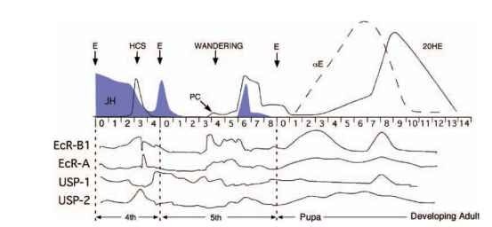
In the Holometabola, complete metamorphosis requires an intervening pupal stage for re-modeling of the larva into an adult. During the last larval instar of Manduca, ecdysteroids rise on two occasions, first in the absence of JH, and later in its presence (Fig. 3). The first ecdysteroid pulse is a small one during days 3-4, referred to as the pupal commitment peak. This is the first time in the life history of the animal that a peak of 20HE occurs in the absence of JH. This triggers cessation of feeding and a wandering behavior aimed at locating a suitable site for pupation. The pupal commitment peak prepares the genome for its response to the next ecdysteroid peak. Although the second pulse of ecdysteroids occurs in the presence of JH, the commitment peak has changed the response of epidermal cells from a larval to a pupal secretory program. Similarly, imaginal discs respond to this peak by differentiating into adult tissues, something not observed in the previous larval stages.
In the Lepidoptera, the new hormonal milieu that triggers metamorphosis produces striking changes in the CNS and musculature. These tissues must be drastically altered during construction of the adult body form. Simultaneously, undifferentiated cells in imaginal discs proliferate and differentiate. For these tissues, the pupal commitment peak sets the stage for three types of cellular responses to the subsequent ecdysteroid peak: programmed cell death (apopto-sis), cellular re-modeling, or differentiation of imaginal discs. For example, some motoneurons that innervate larval-specific structures such as prolegs die shortly after pupal ecdysis. Others persist because of their involvement in the motor patterns involved in adult eclosion, and die soon thereafter. Most larval neurons survive, but are re-modeled to play roles in adult behavior.
The precise mechanisms governing cellular responses to the pupal commitment peak remain obscure, but the identification of EcR and USP subtypes has allowed monitoring of their expression during metamorphosis. For example, epidermal cells of Manduca

FIGURE 3 Hormonal regulation of development in postembryonic stages of the moth, Manduca sexta, exemplifying the Holometabola. During larval development, which extends through five instars, elevated JH levels maintain the “status quo” of immature phenotype. However during the fifth instar, JH levels drop and metamorphosis is triggered by occurrence of a small ecdysteroid peak in the absence of JH. This “pupal commitment” (PC) peak results in a switch from feeding to wandering behavior, and a change in the way tissues respond to subsequent ecdysteroid peaks. The ecdysteroid peak in the presence of JH on day 6 programs the pupal phenotype. During the pupal stage, aE and 20HE again rise in the absence of JH, signaling adult commitment. Fluctuating levels of ecdysteroid receptor mRNA transcripts encoding EcR-B1, EcR-A, USP-1, and USP-2) are shown below. (Adapted from Riddiford, Cherbas and Truman, 2000 and Baker etal., 1987.) respond to the ecdysteroid peak during days 2-3 of the fourth instar by upregulating EcR-B1, not responding to EcR-A, and downregu-lating USP-1 (Fig. 3 ) . However, during the fifth instar, the pupal commitment peak is correlated with sharply increased EcR-B1 and EcR-A expression, and an altered USP-1 response. These altered patterns of expression apparently encode a change in downstream gene expression, leading to a pupal phenotype as well as imaginal disc differentiation during this stage.
During the pupal stage, ecdysteroid levels rise in the complete absence of JH (Fig. 3). This signals commitment to the adult phenotype and accelerated development of imaginal discs. It is remarkable that aE begins to rise on day 1 of the pupal stage, well before elevation of 20HE, and this is correlated with increases in both EcR-B1 and EcR-A expression. This suggests that aE may have a hormonal role itself in programming of the adult stage. Elevation of 20HE occurs in two phases, one beginning on day 3 and a second, steeper rise on day 7. The slow gradual rise coincides with the adult commitment phase, whereas the steeper rise beginning on day 7 elevation is associated with differentiation of new tissues. This latter phase is a rapid rise of EcR-B1 and USP-1 expression.
The hormonal signaling mechanisms governing metamorphosis are complex, and include a diversity of hormones, receptors, and varying temporal patterns of hormone release and receptor expression. Ecdysteroid signaling in the presence or absence of JH can set the stage for changes in the programming of target tissues, such as epidermal cells that secrete cuticle. Depending on the responses of EcR and USP subtypes, qualitatively different cellular programs are initiated.
