Introduction
Assessment of sex is one of the most vital determinations to make when it is necessary to establish identity from skeletal remains. In the first place, this classification effectively cuts the number of possible matches in half, and proper identification could never be made if officials were told to look for a male when the remains were actually those of a female. Moreover, identification techniques such as facial reconstruction would be impossible if sex could not be correctly established. Thus, isolating, interpreting and quantifying the manifestations of sex is an essential part of all skeletal analyses. Unfortunately, this is often not a simple process since male and female attributes span a continuum of morphologic configurations and metric measures in the skeleton. Although some bones are better indicators than others, there is no skeletal feature that rivals the definitiveness of differences between fleshed individuals.
In some cases, remains of clothing, hair, jewelry and other artifacts may be helpful to corroborate a skeletal assessment and indeed lead to a positive identification of the items in question, but they may also be misleading. Today, long hair is not always a female prerogative, nor is a man’s shirt only worn by males. A woman may be wearing her boyfriend’s ring and both sexes often wear earrings. Cross-dressers are also a problem and identification may be delayed or even impossible without a thorough skeletal examination. One must be particularly careful in mass disasters where both bones and personal items may be comingled.
Sexual dimorphism
In normal, living humans, sex is a discrete trait determined by the actions of the XX or XY genotype and diagnosed by observing one of only two possible morphological features. These differences (e.g. external genitalia) are apparent at birth and clearly recognizable even during the prenatal period. In the skeleton, however, the most effective sex indicators do not begin to develop until adolescence, and some are not fully expressed until adulthood. Although almost every human bone has been analyzed, both metrically and morphologically, even a complete, mature pelvis cannot consistently guarantee more than 95% separation of the sexes.
There are two methodological approaches to sexing skeletal remains: morphological and osteometric. Morphologic techniques focus on shape – the bony configurations that are macroscopically visible and differ between males and females. There are important advantages to this approach, especially when a particular form is recognizable despite temporal and population variation. Obvious morphological differences such as the subpubic angle of the pelvis and ramus flexure in the mandible allow optimal separation of the sexes (approaching 95% accuracy). One reason these are so effective is that they are developmental in nature. Subpubic angle expansion is one of several pelvic features that develop during adolescence to accommodate the demands of childbearing on the female pelvis. Ramus flexure results from postadolescent growth in males, whereas the lack of it maintains the straight juvenile form in females. The main drawback of the morphologic approach in general (especially when judgement of size is involved) is that it is based on ‘eye balling’ so if the formation in question is not patently obvious, experience becomes an essential component. The observer must develop a sense of what is relatively large or small, angled or curved, wide or narrow.
Unfortunately, not all skeletal components have consistent and discernible morphologic evidence of sexual dimorphism. Problems arise when differences are only size based and there is no clear male or female shape. Many bony sites are known to exhibit visible differences in size, but osteologists can only determine if they fall into the male or female range after years of experience. However, the overlap between the sexes, along with significant population variation, makes this problematic for all but extreme cases. Although the size of the mastoid process, for example, is often used for sexing, it is difficult to judge by simply looking at it, even if one is familiar with the range of variability within a given group, e.g. Southern African Blacks have much smaller mastoids than Whites or American Blacks. Therefore, metric analysis, based on bone dimensions, is the method of choice for skeletal parts like long bones that do not exhibit clearly definable shape variants. Often a combination of measurements are selected from each bone to maximize sex diagnosis using discriminant function statistics. The major problem with this technique is that standards are temporally sensitive and population specific (both between and within groups) – a formula for Whites may not work on Blacks or Asian Whites, and formulas for American Blacks may not be effective on South African Blacks. Another drawback is the metric overlap of the sexes which can be as high as 85%. Finally, although the metric approach draws heavily on the fact that males tend to be more robust because normal testosterone levels produce greater muscle mass, cultural variation in functional demands may lead to both differences in the expression of sexual dimorphism and its magnitude. This is of particular concern in bones where dimorphism is not primarily developmental, i.e. those that reflect weight bearing and labor-intensive stress.
The diagnosis of sex from the skeleton is further complicated by a number of diverse factors ranging from environmental influences and pathologic conditions to temporal change, diet and occupational stress. In addition, recent genetic and endocrine studies have demonstrated that the expression of the sex genotype can be modified by a malfunction of the appropriate hormonal triggers or on-site receptors. As noted before, all skeletal sites and traits are not equally effective at distinguishing one sex from the other. So the question arises – why bother with the less definitive parts? The answer is simple – skeletal remains are rarely complete and undamaged, even in recent forensic cases. Therefore, the investigator must be able to glean as much information as possible from any bone or fragment. This article presents an overview of current methods and their accuracy for sex determination from the human skeleton.
The Immature Skeleton
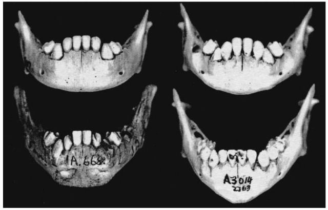
Because sex differences in the immature skeleton are not readily observable before puberty, the few attempts that have been made to look for dimorphic indicators have met with limited success, especially for individuals in the first few years of life. One exception is a recent study of the development of sexual dimorphism in the mandible that revealed differences in shape of the inferior symphyseal border and corpus beginning with the eruption of the central incisors at about seven months until about 4-5 years. As can be seen at the top of Fig.1, some mandibles had a gradually curved or rounded border – these turned out to be female. In contrast, the anterior aspect of the male mandibles (below) extends abruptly downward coming to a point or squaring off at the symphysis. These mandibles clearly demonstrate the sharply angled transition to the lateral body in males so different from the gradual transition in the female. Mandibular corpus (body) shape (not to be confused with dental arcade form) is another important feature. The female is characterized by a distinctly rounded contour of the body resulting from a gradual transition from front to sides, in contrast to the male in whom the sides diverge in a sharp, angular fashion from a nearly horizontal anterior region just past the canines. Even when the male symphyseal base is more rounded, there is still a precipitous descent from the lateral body. Accuracy which averages 81% is higher than that attainable fromadult chin shapes or any other part of the immature skeleton in this age range. By 6-8 years of age, traditional adult chin morphology (squared or undercut in males; rounded in females) was recognizable. The anterior mandible was found to be nearly identical to adults in both form and size by as early as 13. The 69% predictive accuracy in 6-19 year olds was not significantly different from the 71% obtained in adults aged 20 and older.
Attempts to capitalize on sex differences in the size of both the deciduous and adult dentition have not been successful. Recent work has demonstrated that tooth dimensions (length and breadth) do not yield a statistically significant separation of the sexes. Another study attempted to determine sex in the fetal, newborn and 6-month-old ilium. Based on observations that the auricular surface was elevated in males accuracy was reported to be 75% in females and 92% in males in this age group. Metrically, however, no sex difference was found in various dimensions of the ilium in this age group. Sexual differentiation of the pelvis is not clearly defined until adolescence. As detailed below, expected variation in the shape of the pubic bone, sciatic notch and subpubic angle are manifest by the mid-teens in normally developing individuals.

Figure 1 Sex differences in the immature mandible. Female morphology (above) has a rounded corpus shape with a gradual transition from the lateral body to the symphysis. Males (below) show a steep abrupt transition with an angular corpus (not dental arcade) shape
The Adult Skeleton: Morphology
When the morphologic expressions of sexual dimorphismin the adult skeleton are easily distinguishable they provide the most consistently reliable means of diagnosing sex. The best places to look for these formations are the skull and pelvis.
The skull
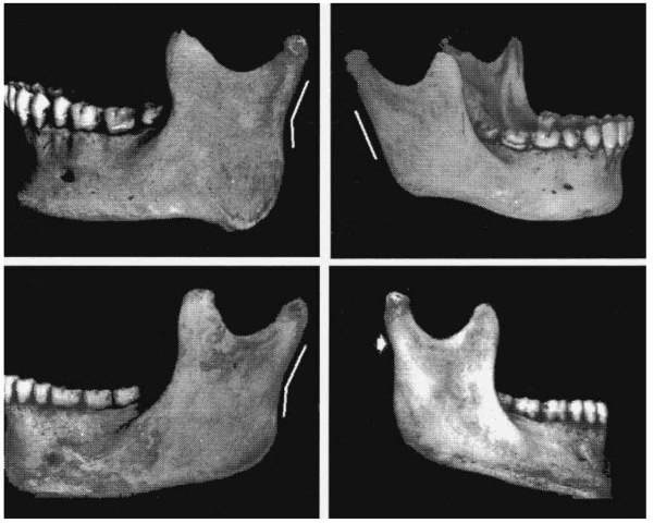
Sex differences have been investigated in nearly every feature of the skull. The most dimorphic part of the skull is the mandible. Fig.2 illustrates sex-linked morphologic configurations of the mandibular ramus. In adult males, ramus shape is characterized by a distinct rearward angulation of the posterior border of the ramus at the level of the occlusal surface of the molars (Fig. 2); this is referred to as ‘ramus flexure’. Females, however, are not flexed at this part of the bone – the posterior border maintains the straight juvenile shape, or, in some females, angulation occurs at a higher point at the neck of the condyle (Fig. 2). This single trait has demonstrated diagnostic accuracy of 91-99% with confirmation by independent tests.
If only the symphyseal region of the mandible is present, a squared or undercut base is often associated with males and a round or pointed inferior outline with females. In modern Whites and Blacks there is an overlap of the female shapes in the males and accuracy is only about 70%. Research has demonstrated that adult chin shapes are highly heritable and only coin-cidentally linked to sex. The same appears to be true for gonial eversion, except that this condition does not even attain separation at levels greater than chance (49%). A better assessment can be made from mandibular condyle shape (68%). Large, thick, oblong condyles are associated with males, and small, round or thin, oblong shapes are usually female.
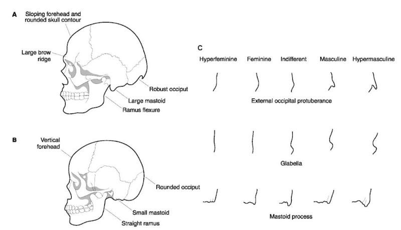
Table 1 contains a list of dimorphic cranial characteristics (Fig.3). In general, the male skull is expected to have more rugged sites of muscle attachments, a prominent glabellar region, and thicker bones. A large, pronounced supraorbital torus and rugged occipital protuberance can be considered reliable male characteristics. When viewed laterally, the sloping male forehead gives the vault a more rounded outline than that of the relatively vertical female forehead. Males have larger mastoid processes. Both frontal and parietal eminences tend to be better developed in females. Some differences in the cranium are of a proportional nature such as the orbits. Because eyes are of similar size in both sexes, the orbits appear to take up a larger portion of the face in females. Females also tend to have round or square orbits with sharp superior orbital margins, in contrast to the more rectangular orbits with rounded margins in males.

Figure 2 Sex difference in the adult mandible. The posterior ramus of adult males (left) is flexed at the level of the occlusal surface of the molars. The female ramus maintains the straight juvenile shape (top right) or may flex above the occlusal level near the neck of the condyle (lower right).
Table 1 Morphologic sex differences in skull and pelvis
| Trait | Male | Female |
| General appearance | Rugged | Smooth |
| Supraorbital ridges | Medium to large | Small to absent |
| Mastoid processes | Medium to large | Small to medium |
| Occipital area | Muscle lines and protuberance | Muscle lines and protuberance not marked |
| marked or hooked | ||
| Frontal eminences | Small | Large |
| Parietal eminences | Small | Large |
| Orbits | Rectangular | Rounded |
| Small relative to face | Large relative to face | |
| Rounded margins | Sharp margins | |
| Forehead | Sloped | Vertical |
| Mandibular ramus flexure | Ramus flexure | Straight ramus |
| Palate | Larger, broader, tends to U-shape | Small, tends to parabola |
| Teeth | Large, lower M1 more often 5 cusped | Small, molars often 4 cusped |
| Pubic symphysis | Higher | Lower |
| Pubic bone | Triangular | Square or rectangle |
| Subpubic angle | Narrow V-shaped, acute angle | Wide U-shaped, obtuse angle |
| Acetabulum | Large, tends to be directed laterally | Small, tends to be directed anterolaterally |
| Greater sciatic notch | Narrow, deep | Wide, shallow |
| Ischiopubic rami | Rough everted margin | Gracile, narrows near symphysis |
| Sacroiliac joint | Large | Small, oblique |
| Postauricular space | Narrow | Wide |
| Preauricular sulcus | Rarely present | Often present, well developed |
| Iliac tuberosity | Large, not pointed | Small or absent, pointed or varied |
| Sacrum | Longer, narrower with more evenly | Shorter, broader, with tendency to marked |
| distributed curvature; often 5 or more segments | curvature at S1-2S2-5; 5 segments the rule | |
| Pelvic brim or inlet | Heart-shaped | Circular, elliptical |

Figure 3 Sex differences in the skull (see Table 1). (A) Male features; (B) female features; (C) morphologic gradations.
In all skeletal assessments, it is important to remember that humans vary both temporally and spatially. To this one may add variation arising from growth factors, environmental stress and pathology or trauma. Therefore, small or absent brow ridges or prominent eminences are not necessarily female indicators. The same can be said of a rounded chin and a less rugged occipital region, both of which are not uncommonly seen in males. In this regard, population differences are a major cause of variability in the expression of sexual dimorphism.
The pelvis
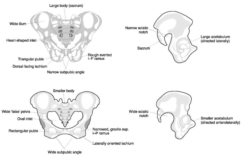
The pelvic girdle is composed of three bones: two coxal (or hip) bones and the sacrum(Fig.4). Each hip bone is in turn the product of the fusion of three bones: the ilium, ischium and pubis. As mentioned earlier, the female pelvis must be designed to accommodate childbirth whereas the only requirements for males are support and locomotion. These differences are manifest in various parts of this complex structure. Table 1 summarizes the morphologic differences in the pelvis.
The most easily assessable morphologic feature is the subpubic angle formed by the ventral articulation of the pubic bones at the symphysis (Fig. 4). The male is characterized by roughly triangular pubic bones that forma narrow subpubic angle. Pubertal growth modifications in females are centered on the facilitation of childbirth. Elongation of the pubic bones creates a more rectangular shape and results in a much wider subpubic angle and larger outlet. Further enlargement of the pelvic outlet is accomplished by widening of the greater sciatic notch in females, but this feature may show considerable variability and is sometimes difficult to assess. If the sacrum can be articulated, it gives a much clearer picture of the width of the notch by completing the dorsal portion. Other female adaptations which can be used in sex determination include a rounded (as opposed to the heart-shaped male) inlet, a dorsally oriented tilt of the sacrum, a small, and everted ischial spine. The upper section or ‘false pelvis’ is broader and deeper in males whereas the lower portion or ‘true pelvis’ is wider in females. The ischial tuberosity is positioned more dorsally in males and laterally in females. A uniquely male trait is the rough everted area of the medial border of the ischiopubic ramus for the attachment of the crus (corpus cavernosum) of the penis. This ramus is more gracile in females and narrows as it approaches the symphysis. In the sacrum, females show a proportionally smaller superior articular surface relative to the total width of the wings.
Figure 4 Sex differences in the pelvis (see Table 2). Male above, female below.
As in all skeletal remains, the most diagnostic parts are not always available, especially following extended periods of exposure to the elements or years of burial. Since spongy bones are more prone to destruction, highly dimorphic bones like the pubis may not be available for diagnosis. Other clues to sex can be found in the sacroiliac joint and relate to its greater mobility in females. In many individuals there are accessory articular facets in males on both the iliac tuberosity and dorsal sacrum. If the sacrum is fused to the ilium, the individual was most likely male, in the absence of trauma or disease.
Other postcranial indicators
If the most reliable sites are missing, at least some indication of sex can also be found in certain features of the long bones and ribs. A foramen in the olecra-non fossa of the humerus has been linked to females. If a long bone is large and robust with pronounced muscle attachments these male indicators take precedence. Rib morphology also shows sex differences. If the sternal end has large projections at the superior/ inferior borders, this is a male characteristic. Post-menopausal females may also exhibit this phenomenon, but the projections are much thinner. It must be kept in mind that these fragile projections are often lost or broken. If the costochondral junction has bony projections extending from the floor of the pit, the individual is likely to be female.
The Adult Skeleton: Metric Analysis
Osteometric analysis can also yield high levels of accuracy for the diagnosis of sex. Techniques range from the calculation of a simple sectioning point derived from a single measurement to complex multi-variate discriminant function analysis. Indices formed by the relationship of one dimension to another like that of the ischiopubic index allow male/female comparisons while eliminating size as a factor. This is important since the sexes can have as much as an 85% overlap for size alone.
Discriminant function analysis is one of the most commonly used techniques to develop sex determination formulas using one or more measurement from the skeleton. In general selection of dimensions for a formula depends on levels of intercorrelation as well as the degree of difference between the sexes. It is, for example, very likely that femoral distal breadth is significantly correlated with tibial proximal breadth and therefore one of these may suffice to provide the best result. In long bones it has been observed that epiphyseal measurements are better indicators of sex than length or diaphyseal dimensions. It has also been shown that, metrically, sexual dimorphism in the long bones is more diagnostic than in the skull and necessitates the use of fewer dimensions. For example, one may need several skull measurements to obtain a sexing accuracy as high as that of the femoral or humeral head diameter alone.
With this in mind, univariate discriminant functions were calculated using eight single dimensions fromthe humerus, femur and tibia (Table 2). Although most of the dimensions used are standard measurements (clearly defined in major reference topics) these dimensions were also selected because these segments of the skeleton are commonly present at a crime scene or grave even if the skeleton is badly fragmented. In order to make this methodology applicable to diverse populations, osteometric analyses of modern (twentieth century) skeletal samples have been conducted on US Whites and Blacks, South African Whites and Blacks, and Asians including mainland Chinese and Japanese. Table 2 lists descriptive statistics for males and females along with the average of the two sexes (sectioning point) and percentage accuracy. In all samples, males were significantly larger than females. Using this technique, determination of sex is accomplished by comparing the dimension of an unknown bone with the sectioning point for that population. For example, if the humeral head diameter of an unknown American White is 47 mm, classification would be ‘male’ (any value larger than 45.6 mm classifies a person as male in this group). Although overall accuracy is 84.8% (Table 2), the farther the measurement is from the sectioning point, the greater the likelihood of correct sexing. Moreover, if the humeral head diameter is greater than the male mean (48.6 mm) or less than the female mean (42.6 mm), the probability of correct determination would be much higher. It must be emphasized however, thatitis necessary to knowracial affinityinthese cases unless the values of the unknown specimen are extreme.
As noted earlier, metrics are group specific. As can be seen in these figures, populations differ fromeach other in the degree and range of dimorphism exhibited by various dimensions. This reflects differences in both size and proportions. In general, South African Whites are the largest of the groups assessed whereas the average South African Black is among the smallest with a mean closer to that of the Chinese and Japanese. Table 2 also reveals population differences in the locus of dimorphism. For example, femoral distal breadth is the most diagnostic measurement in South African Whites, whereas maximum dimorphism in the Japanese is found in the epicondylar breadth of the humerus.
Table 2 Metric determination of sex and classification accuracy from single long bone dimensions (mm) in American Whites and Blacks, South African Whites and Blacks, and Chinese and Japanese.
| Terry Whites | Terry Blacks | S Afr Whites | S Afr Blacks | Chinese | Japanese | ||||||
| Dimensions | Mean SD | Mean | SD | Mean | SD | Mean | SD | Mean | SD | Mean | SD |
| Male: 46 | Male: 45 | Male: 44 | Male: 37 | Male: 33 | Male: 39 | ||||||
| n | Female: 45 | Female: 44 | Female: 42Female: 44 | Female: 29 | Female: 32 | ||||||
| Humeral head diameter | |||||||||||
| Males | 48.6 4.18 | 48.0 | 3.17 | 49.0 | 3.28 | 43.8 | 2.23 | 45.1 | 2.56 | 44.0 | 1.72 |
| Females | 42.4 2.33 | 42.8 | 2.29 | 43.0 | 2.28 | 37.7 | 2.08 | 39.3 | 2.20 | 39.1 | 2.67 |
| Average | 45.6 4.60 | 45.4 | 3.81 | 46.1 | 4.13 | 40.5 | 3.73 | 42.4 | 3.76 | 41.8 | 3.27 |
| % Accuracy | 84.8 | 86.8 | 84.6 | 90.9 | 80.5 | 87.3 | |||||
| Humeral epicondylar breadth | |||||||||||
| Males | 63.9 4.35 | 64.5 | 4.4263.6 | 3.67 | 61.3 | 6.43 | 58.5 | 3.87 | 59.9 | 2.11 | |
| Females | 56.3 3.52 | 57.5 | 3.02 | 55.7 | 2.69 | 53.5 | 3.62 | 52.2 | 2.75 | 52.2 | 3.95 |
| Average | 60.25.50 | 61.0 | 5.13 | 59.7 | 5.10 | 57.0 | 10.05 | 55.6 | 4.6256.4 | 4.91 | |
| % Accuracy | 83.7 | 86.8 | 88.4 | 88.6 | 84.4 | 91.1 | |||||
| Femoral head diameter | |||||||||||
| Males | 48.9 3.14 | 48.1 | 3.23 | 48.4 | 2.76 | 45.3 | 2.33 | 46.2 | 2.60 | 46.0 | 1.86 |
| Females | 42.8 2.39 | 42.6 | 2.03 | 43.1 | 2.13 | 39.2 | 2.60 | 41.0 | 2.31 | 40.8 | 2.54 |
| Average | 45.9 4.13 | 45.4 | 3.86 | 45.8 | 3.64 | 42.0 | 3.94 | 43.8 | 3.58 | 43.6 | 3.40 |
| % Accuracy | 82.6 | 88.0 | 85.9 | 91.9 | 83.1 | 88.6 | |||||
| Femoral midshft circumference | |||||||||||
| Males | 91.5 4.82 | 92.7 | 5.65 | 92.0 | 5.47 | 90.0 | 5.16 | 85.3 | 6.37 | 85.4 | 4.16 |
| Females | 82.8 5.67 | 84.0 | 5.73 | 84.5 | 5.14 | 78.8 | 4.74 | 75.3 | 4.66 | 78.3 | 6.40 |
| Average | 87.26.80 | 88.4 | 7.16 | 88.3 | 6.48 | 84.0 | 7.45 | 80.6 | 7.53 | 82.2 | 6.32 |
| % Accuracy | 79.1 | 79.1 | 74.3 | 89.9 | 81.7 | 78.5 | |||||
| Femoral distal breadth | |||||||||||
| Males | 83.1 4.44 | 82.8 | 4.68 | 84.0 | 4.61 | 79.2 | 4.24 | 80.2 | 4.47 | 81.1 | 2.73 |
| Females | 75.3 3.58 | 74.7 | 3.66 | 75.2 | 3.27 | 69.6 | 4.78 | 69.8 | 3.00 | 72.4 | 4.31 |
| Average | 79.25.6278.8 | 5.85 | 79.7 | 5.98 | 74.0 | 6.60 | 75.3 | 6.50 | 77.1 | 5.58 | |
| % Accuracy | 79.4 | 87.0 | 90.5 | 86.4 | 92.3 | 85.5 | |||||
| Tibial proximal breadth | |||||||||||
| Males | 76.0 3.68 | 77.1 | 4.14 | 78.24.43 | 74.9 | 3.72 | 72.8 | 3.74 | 73.5 | 2.87 | |
| Females | 68.6 3.69 | 68.3 | 3.00 | 69.8 | 2.96 | 64.9 | 7.37 | 63.6 | 3.36 | 66.2 | 4.42 |
| Average | 72.3 5.25 | 72.8 | 5.68 | 74.1 | 5.63 | 69.5 | 7.80 | 68.5 | 5.81 | 70.2 | 5.16 |
| % Accuracy | 85.9 | 89.1 | 87.7 | 86.5 | 89.5 | 88.6 | |||||
| Tibial circumference at nutrient foramen | |||||||||||
| Males | 96.25.76 | 100.1 | 6.91 | 97.3 | 6.13 | 98.4 | 6.35 | 93.1 | 6.5291.6 | 4.90 | |
| Females | 86.7 7.90 | 90.0 | 6.20 | 87.2 | 6.13 | 85.1 | 5.20 | 76.4 | 5.77 | 82.5 | 8.56 |
| Average | 91.5 8.36 | 95.1 | 8.28 | 92.4 | 7.94 | 91.2 | 8.78 | 85.3 | 10.41 | 87.5 | 8.13 |
| % Accuracy | 76.1 | 80.4 | 82.1 | 83.50 | 90.4 | 80.0 | |||||
| Tibial distal breadth | |||||||||||
| Males | 47.7 3.15 | 47.6 | 3.76 | 46.8 | 2.62 | 45.4 | 2.56 | 45.1 | 2.38 | 45.3 | 2.18 |
| Females | 43.3 2.86 | 47.6 | 2.70 | 41.9 | 2.50 | 39.9 | 2.23 | 39.0 | 2.34 | 40.7 | 2.41 |
| Average | 45.5 3.71 | 45.4 | 3.98 | 44.4 | 3.53 | 42.4 | 3.63 | 42.2 | 3.87 | 43.3 | 3.24 |
| % Accuracy | 78.3 | 80.4 | 80.7 | 87.5 | 90.1 | 82.5 | |||||
Parturition Scars
In the last 30 years, there has been considerable debate as to whether one can determine if pregnancy and parturition leave traces in the skeleton. The initial hypothesis was that these processes can tear the ligaments of the pubic bones and sacroiliac joint and are responsible for causing scars or pits at these sites. These manifestations are found on the dorsal surface of the pubis and preauricular sulcus at the inferior margin of the auricular surface of the ilium. It was also claimed that the number of births can be estimated by the size and number of so-called ‘parturition pits’. However, more recent studies using bones from females of known parity disagreed with this hypothesis. The scars were found in many females who had never had children, and scar size and shape showed variation; these, too, were not related to the number of children borne by the woman. Obviously, other factors must cause similar scarring. These may include horseback riding, habitual squatting, and exercise involving hopping and jumping.
Summary and Conclusions
Although there are many techniques to determine sex from practically every part of the skeleton, there are a number of caveats. The accuracy of both morphological and osteometric sex assessment depends on rigorous training and first-hand experience in observing skeletal remains. The overlap between the sexes implies that structures show gradations of differences within and between sexes and populations. This gradation must be carefully evaluated by studying large documented skeletal collections from different regions of the world. Bone measurement is a tedious process and requires consistency and standardization. The technique has a long history and although there is international agreement among scientists on how measurements are taken (to allow comparability), individual variation in measuring and interobserver error are always troublesome factors.
Even though decades of research have resulted in a plethora of methods and standards of assessment, many factors complicate the determination of sex from the skeleton. Age must be considered because the skeleton does not exhibit maximum dimorphism until sexual maturation and growth are complete. Problems also arise when one only has a partial or fragmented skeleton. It is not always easy to distinguish certain bones of a male of 17 from one in his 20s and since growth can continue into the latter decade, sex differences may neither be clear metrically nor morphologically. This is further complicated by the fact that there is significant variation between populations within each race who inhabit different geographic regions and constitute established gene pools.
Finally, since anything that affects the growth, development, maintenance and remodeling of the skeleton can modify the expression of sexual dimorphism, trauma and disease can result in mis-sexing. Some conditions cause abnormal bony enlargement whereas others result in atrophy or obliterate normally clear morphologic differences. The intimate relationship between bone and muscle means that even in the absence of obvious damage to a skeletal component, muscular impairment can lead to inappropriate remodeling. All of these factors, above and beyond the normally occurring variability, subtleness, and overlap of features in the human skeleton, make it essential that only properly trained and experienced forensic anthropologists be consulted in every case involving the medicolegal system.

