Introduction
There are many instances of human remains where the only item available for identification is a denuded skull. How useful it would be, to be able to replace the missing soft tissues of the face and to see what the individual looked like in life. This is, in fact, the goal of forensic facial reconstruction (FFR), namely, to replace an artificially rendered version of the facial soft tissue over the unidentified skull, in such a way as to suggest the identity of the deceased. It has been suggested that the word (facial) reconstruction, be replaced by the term reproduction or approximation. For the purposes of this article the more widely used term, reconstruction, will be used.
In forensic science, facial soft-tissue depth measurements are of use in the following instances:
• Assisting in the alignment of a face image to a skull image in a technique called skull-photo superim-position. This is an estimation of ‘fit’ between two known images.
• Two-dimensional facial reconstruction where a soft-tissue profile is calculated, drawn and adapted to a tracing of the skull profile.
• Computerized application of a reconstructed or stored face image to a found skull.
• Three-dimensional rendering of the face, in clay or other malleable material.
The chief thrust of this article is to discuss the amount and form of facial soft tissue and the processes involved in three-dimensional FFR.
At the outset we should grasp the limits of FFR. What it is not able to do is to reproduce the exact features of an unknown face. What is possible, however, is that it can produce a semblance of the victim’s appearance that could just be of assistance in the identification process.
Concept of Facial Reconstruction
How can one learn more about facial soft-tissue architecture? Certainly, open dissection is a good start. In fact, a practical understanding of head and neck anatomy should be a prerequisite for all those intending to work in FFR. The Further Reading list at the end of this article refers to in-depth surveys of research work done in the field of FFR; comprehensive summaries of major works on forensic anthropology, and reviews on the current status of facial reconstruction.
The earliest tables of soft-tissue depth measurements were drawn up at the end of the nineteenth century (when the faces of notable people such as Kant, Schiller and Bach were reconstructed). Using thin knives and needles, investigators probed and recorded the facial soft-tissue depth of cadavers at a number of landmarks. Only in the third decade of the twentieth century was facial reconstruction used for forensic purposes. Over the years, tables of measurements were established for the three major ethnic groups:Caucasoid, Mongoloid and Negroid. The Negroid Group was composed of African-Americans. These are a blend of Negroid, Caucasoid and Mongoloid types. Although no ethnic group is, correctly speaking, biologically homogenous, there are certain isolated groups that have resisted crossgroup intermixture for long periods. In order to obtain a more homologous set of measurements for the Negroid, a table of facial soft-tissue depths for the African Zulu male was published in 1993. The data derived from the study are linked with the production of midline and oblique face profiles and are best understood in their entirety in the original work. As is the case in this last study, more researchers are now measuring tissue thickness using cephalometric radiography, ultrasonography and, more recently, computed tomography and magnetic resonance imaging. Various facial tissue depth measurements are also available for Caucasoid children, though these are representative of humans in their most rapidly changing stage of life.
In order to compare the measurements of the different studies, soft-tissue measurements should be taken at the same landmarks in each study. The most commonly used landmarks are illustrated in Fig. 1. Although the Mongoloid landmarks are fewer in number, they correspond to those used on white and black studies (see Table 2). There are also a number of smaller scattered studies on Caucasoid and Negroid groups, listed in the review mentioned in the Further Reading list.
Of course the value of any measurement is limited to a single locus and offers no information on the depth or character of the adjacent tissue. This means the person doing the modeling (the ‘sculptor’) begins work with a very limited amount of information indeed.
There is some disagreement as to who should carry out the practical work of FFR. It seems the ideal situation should occur when the attributes of scientist and artist exist in the same worker, but this is rare. Usually the anthropologist works together with an artist to model the face. Some feel it unnecessary to use trained artists or sculptors, as the outcome of the work can be negatively affected by their creative tendency. This could well be the case if the artist is given free reign for interpretation. On the other hand, if ‘trained’ scientific experts are used, they need to have a fair modicum of manual dexterity and modeling skill to enable them to reconstruct a realistic-looking face. The ideal kind of worker is a professionally trained realist sculptor, such as a portrait artist, who can honestly render a three-dimensional face that looks like a face. In all cases, artistic ability should be respectful of, and subservient to, the dictates of science. In short, there is a distinct need for artistic talent, but not for artistic license.
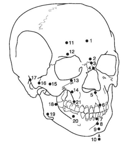
Figure 1 Landmarks for soft-tissue depth measurements. See Table 1for landmark names corresponding to the numbers on the illustration.
There are fundamentally two different approaches to reconstructing the human face. The first makes use of objectivity, measurements and rules. The second, made popular by Russian workers, is based on anatomical knowledge, critical observation and subjective evaluation. The two methods could be called morphometric and morphoscopic, respectively. One might be forgiven for feeling that the morphoscopic method is unscientific and is therefore best left alone; however, there have been a number of talented workers who have used it with success. The morphoscopic method will be only briefly mentioned, as the main focus will be placed on the morphometric approach to facial reconstruction.
In order to make FFR a scientifically acceptable technique, an objective method must be devised that can produce the same result each time. Theoretically, if three sculptors are supplied with identical information and copies of a skull, they should be able, using a standardized and controlled technique, to reconstruct three similar faces. If this can be accomplished, a reliable and repeatable method of facial reconstruction will have been produced, which will have the added advantage of being granted weight in the eyes of the law.
With this in mind the technique should conform to certain regulations. Firstly, only standard landmarks may be used. Secondly, only average soft-tissue measurements may be employed. Thirdly, the areas between landmarks must be filled in with mathematically graded clay strips and filler material. Finally, the sculptor must make use of ‘rules of thumb’ and controlled modeling techniques. Admittedly, the result may be a rather bland approximation that relies on the general form of the skull for any suggestion of likeness. The paradox is that people are usually remembered for their differences from the average; that is, for their peculiarities.
Practical Process
In all cases, the sculptor should be provided with as much information on the deceased as possible i.e. ethnic group, age, gender, stature and weight.
In most cases the mandible is attached to the skull in what is called centric occlusion:with upper and lower teeth together. Preferably, the mandible should be placed in ‘centric relation’, with a freeway space between the teeth. This represent a jaw that is relaxed. People do not normally walk around with their teeth clenched.
There are differing opinions as to whether the reconstruction should be carried out on the original skull or whether a duplicate casting should be used. If a casting is made, it is well to bear in mind that distortion can occur during the impression-taking process. Also, if the copy is cast in plaster of Paris a uniform dimensional change could take place during the setting phase. The thoroughly cleaned skull (or casting) is then mounted on a stand, ready for the modeling process.
Morphoscopic method
In this method, the face is considered as being individually highly characteristic. The skull is first examined for any ‘clues’ that could suggest the amount of muscle development, for example the size and shape of the mastoid process or the degree of eversion at the angle of the mandible. Reconstruction begins with modeling three of the main muscles of mastication: the temporalis, masseter and the buccinator (some will disagree that the last named is a muscle of mastication). Care should be taken not to exaggerate the bulk of these muscles. Though powerful, they are not all that thick. For example, a fair amount of the volume of tissue overlying the ramus is made up of fat and parotid gland, which gives the side of the jaw a characteristic shape. Likewise, the buccinator, though plump in children (as an aid to suckling) is not at all that thick in adults. The pterygoid muscles do not play a role in defining the form of the face.
Next, the circular muscles around the mouth and eyes are built up. These are relatively weak muscles and are quite thin and flat except for their junctions with other muscles, for example the commissures at the corners of the mouth. Other tissues to be added are the parotid glands and any fatty deposits, if suspected. Finally a layer of ‘skin’, varying in thickness between 0.5 and 1.0 cm, is adapted to the surface of the reconstruction and textured. The features are then modeled, more by anatomical knowledge, feel and experience, than by rules. Some sculptors use the morphoscopic approach throughout, but every now and again pierce the clay at the landmarks to check for average depth. The stages in the process are illustrated in Fig. 2.
Morphometric method
To begin with there must be some way of indicating the depth of ’tissue’ to build up at the landmarks. Innovative workers have used clay mounds, rubber cylinders, carding strips and matchsticks as depth indicators. In the case of the reconstruction illustrated in Fig. 3, thin balsa wood rods were used.
The rods are cut to lengths matching the data in the appropriate tables of soft-tissue depths (Tables 1 and 2) and stuck to the skull (or cast) at the relevant landmarks. Small, 1.5 cm ‘islands’ of clay are built up to the height of each rod to prevent their accidental displacement. Flattened strips of clay, 1 cm wide and evenly graded in thickness from one depth rod to the next, are placed between the islands. In a similar manner, the voids between islands and strips are filled in, the rods removed, and the surface smoothed off, taking care not to thicken or compress the clay.
Except for the features, the whole skull and face are covered in this manner. The stages in this process are shown in Fig. 3. Drawn guides to the reconstruction of the features are illustrated in Fig. 4, and are explained in the following text.
Eyes Most sculptors insert artificial eyes into the sockets, although some prefer to construct eyeballs from clay, with the irises carved out. The plausible reason for this is that the color of the deceased’s eyes is usually unknown. Perhaps, if the skull is that of a
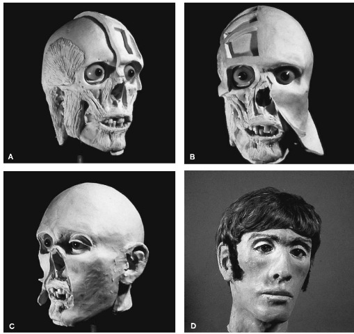
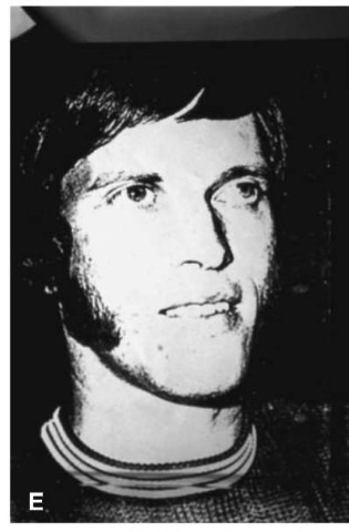
Figure 2 The morphoscopic method of facial reconstruction: (A) modeling the facial muscles and scalp strips; (B) adaptation of ‘skin’ covering; (C) modeling the ears and eyes; (D) the final, painted casting (sideburns added later); (E) a photograph of the identified person.
Negroid or an Asiatic, one could hazard a guess that the irises are dark.
The eyeball is positioned with its corneal surface on a plane created by drawing a midsocket vertical line between the upper and lower margins of the bony orbital rim. The pupil is centered behind the junction of this line, with another crossing it at right angles across the widest part of the orbit.
Generally, the upper eyelid covers the upper third of the iris and the lower rim is positioned at the bottom of the iris. If the ethnicity of the skull is Mongoloid, the eyelids could be slanted upwards and outwards. Otherwise, the slant of the eye can be roughly determined by a line running from the lachrymal opening in the medial wall of the orbit to the tubercle on the lateral orbital rim. In modeling the eyelids, one should take into account the thickness of the inner canthus (2 mm) and outer canthus (4 mm).
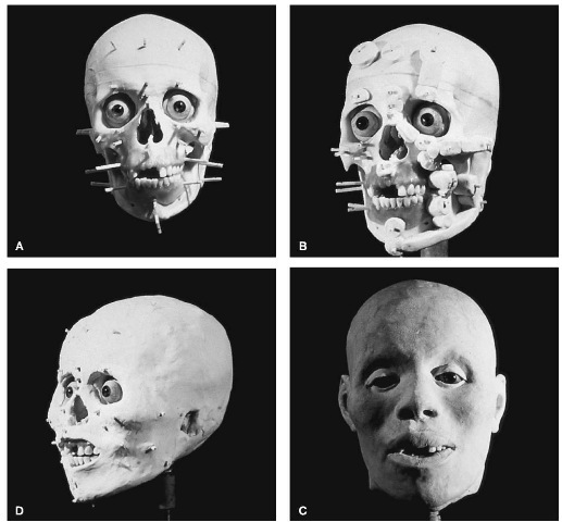
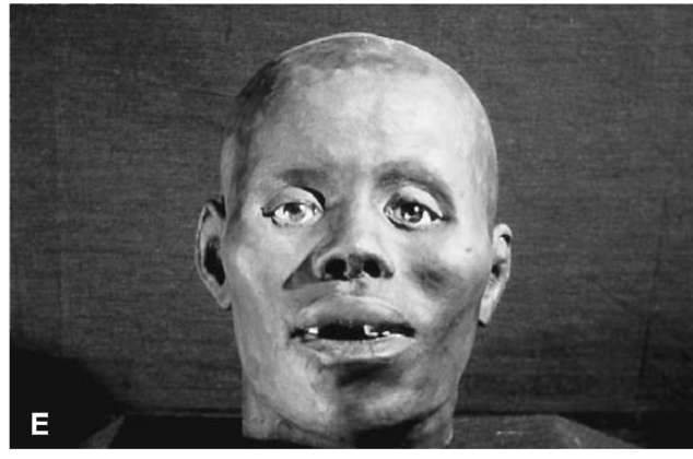
Figure 3 The morphometric method of facial reconstruction: (A) showing depth pegs stuck to skull, at landmarks; (B) islands and graded strips placed between pegs; (C) ‘skin’ surface smoothed off; (D) finished clay representation; (E) final painted casting.
Nose The nose is considered a dominant feature. Yet, if it is within the bounds of average for that particular ethnic group, it is hardly noticed. Only if out of the ordinary is it of any significance in recognition. Except for the nasal bones, there is no underlying bony support for the soft tissues of the nose. Despite this, certain rules have been developed, the main ones being aimed at the length, height and width of the nose.
Table 1 Facial tissue thickness (mm) of American Whites
| Landmark | Emaciated | Normal | Obese | |||
| Male (3) | Female (3) | Male (37) | Female (8) | Male (8) | Female (3) | |
| Midline | ||||||
| 1. Supraglabella | 2.25 | 2.50 | 4.25 | 3.50 | 5.50 | 4.25 |
| 2. Glabella | 2.50 | 4.00 | 5.25 | 4.75 | 7.50 | 7.50 |
| 3. Nasion | 4.25 | 5.25 | 6.50 | 5.50 | 7.50 | 7.00 |
| 4. End of nasals | 2.50 | 2.25 | 3.00 | 2.75 | 3.50 | 4.25 |
| 5. Midphiltrum | 6.25 | 5.00 | 10.00 | 8.50 | 11.00 | 9.00 |
| 6. Upper lip margin | 9.75 | 6.25 | 9.75 | 9.00 | 11.00 | 11.00 |
| 7. Lower lip margin | 9.50 | 8.50 | 11.00 | 10.00 | 12.75 | 12.25 |
| 8. Chin-lip fold | 8.75 | 9.25 | 10.75 | 9.50 | 12.25 | 13.75 |
| 9. Mental eminence | 7.00 | 8.50 | 11.25 | 10.00 | 14.00 | 14.25 |
| 10. Beneath chin | 4.50 | 3.75 | 7.25 | 5.75 | 10.75 | 9.00 |
| Bilateral | ||||||
| 11. Frontal eminence | 3.00 | 2.75 | 4.25 | 3.50 | 5.50 | 5.00 |
| 12. Supraorbital | 6.25 | 5.25 | 8.25 | 7.00 | 10.25 | 10.00 |
| 13. Suborbital | 2.75 | 4.00 | 5.75 | 6.00 | 8.25 | 8.50 |
| 14. Inferior malar | 8.50 | 7.00 | 13.25 | 12.75 | 15.25 | 14.00 |
| 15. Lateral orbit | 5.00 | 6.00 | 10.00 | 10.75 | 13.75 | 14.75 |
| 16. Midzygomatic arch | 3.00 | 3.50 | 7.25 | 7.50 | 11.75 | 13.00 |
| 17. Supraglenoid | 4.25 | 4.25 | 8.50 | 8.00 | 11.25 | 10.50 |
| 18. Occlusal line | 12.00 | 11.00 | 18.25 | 17.00 | 23.50 | 20.25 |
| 19. Gonion | 4.50 | 5.00 | 11.50 | 12.00 | 17.50 | 17.50 |
| 20. Sub M2 | 10.0 | 9.50 | 16.00 | 15.50 | 19.75 | 18.75 |
| 21. Supra M2 | 12.00 | 12.00 | 19.50 | 19.25 | 25.00 | 23.75 |
The nose length and height are determined by drawing a line tangent to the terminal third of the nasal bones in the midline and continuing it downward and forward to meet another line, extending the axis ofthe anterior nasal spine, running upwards and outwards.
There are various rules for the width of the nose:
• It is the same as the inner canthal distance (which in turn is often claimed to be the same as the palpebral fissure length),
• It is 10 mm wider than the bony width in whites, and 16 mm in blacks.
• It is 1.67 times the width of the bony nasal aperture.
• It is one-third larger than the bony width (the pyri-form aperture is three-fifths of the interalar width).
• A well controlled study of 197 skulls and face masks in the Terry collection showed the interalar width to be the pyriform aperture width (PAW) + 12.2 mm in Caucasoid males, and PAW + 16.8 mm in Negroid males.
Measurements of this sort need to be extended over a large number of samples, varying in gender, age and ethnicity, before they are of sound value.
The most characteristic shapes of the nose are the bridge curve (arched, straight, dipped, wide or narrow), much of which is suggested by the form and direction of the nasal bones, and the nose tip which has no bony clue and is highly variable. The alae differ more in size than they do in shape. The nostril shape is another variant. In the high narrow nose the oval-shaped nasal opening is angled more vertically, while in the wide flat nose it is horizontally positioned.
Mouth The mouth is a capricious feature, although in some ethnic groups it is rather characteristic of that group. For example, Negroid lips are almost inevitably everted and are thicker than Caucasoid lips. A rule-of-thumb is that the width of the mouth, or intercommissural distance, is equal to the inter-pupillary distance. Dentists are taught that the corners of the mouth are positioned at the rear edge of the upper canines, with the upper lip covering two-thirds of the upper anterior teeth. In some cases the upper lip reaches down to the incisal edges. The fullness and contour of the lips are dependent on ethnicity, age and dental support. The philtrum or vertical midline gully in the upper lip is also a variable structure. If the upper dental arch is wide, the philtrum is frequently flat. The converse is also often true, especially if the nose is narrow and tipped upwards. The anteroposterior relationship of upper to lower lip is characteristic in cases of an open bite and a jutting lower jaw.
Table 2 Facial tissue thickness (mm) of American Blacks and (Negroids) and Japanese (Mongoloids)
| Landmark | Black | Japanese | |||
| Male | Female | Point | Male | Female | |
| Midline | |||||
| 1. Supraglabella | 4.75 | 4.50 | m | 3.00 | 2.00 |
| 2. Glabella | 6.25 | 6.25 | gl | 3.80 | 3.20 |
| 3. Nasion | 6.00 | 5.75 | n | 4.10 | 3.40 |
| 4. End of nasals | 3.75 | 3.75 | rhi | 2.20 | 1.60 |
| 5. Midphiltrum | 12.25 | 11.25 | - | - | - |
| 6. Upper lip margin | 14.00 | 13.00 | - | - | - |
| 7. Lower lip margin | 15.00 | 15.50 | - | - | - |
| 8. Chin-lip fold | 12.00 | 12.00 | ml | 10.50 | 8.50 |
| 9. Mental eminence | 12.25 | 12.25 | pg | 6.20 | 5.30 |
| 10. Beneath chin | 8.00 | 7.75 | gn | 4.80 | 2.80 |
| Lateral | |||||
| 11. Frontal eminence (L) | 8.25 | 8.00 | - | - | - |
| Frontal eminence (R) | 8.75 | 8.00 | - | - | - |
| 12. Supraorbital (L) | 4.75 | 4.50 | - | - | - |
| Supraorbital (R) | 4.75 | 4.50 | sc | 4.50 | 3.60 |
| 13. Suborbital (L) | 7.50 | 8.50 | - | - | - |
| Suborbital (R) | 7.75 | 8.50 | or | 3.70 | 3.00 |
| 14. Inferior malar (L) | 16.25 | 17.25 | - | - | - |
| Inferior malar (R) | 17.00 | 17.25 | - | - | - |
| 15. Lateral orbit (L) | 13.00 | 14.25 | - | - | - |
| Lateral orbit (R) | 13.25 | 12.75 | ma | 5.40 | 4.70 |
| 16. Zygomatic arch (L) | 8.75 | 9.25 | - | - | - |
| Zygomatic arch (R) | 8.50 | 9.00 | zy | 4.40 | 2.90 |
| 17. Supraglenoid (L) | 11.75 | 12.00 | - | - | - |
| Supraglenoid (R) | 11.75 | 12.25 | - | - | - |
| 18. Occlusal line (L) | 19.50 | 18.25 | - | - | - |
| Occlusal line (R) | 19.00 | 19.25 | - | - | - |
| 19. Gonion (L) | 14.25 | 14.25 | - | - | - |
| Gonion (R) | 14.75 | 14.25 | go | 6.80 | 4.00 |
| 20. Sub M2 (L) | 15.75 | 16.75 | - | - | - |
| Sub M2 (R) | 16.50 | 17.25 | rrH | 14.50 | 12.30 |
| 21. Supra M2 (L) | 22.25 | 20.75 | - | - | - |
| Supra M2 (R) | 22.00 | 21.25 | 10.20 | 9.70 | |
| Sample size | 44 | 15 | 9 | 7 | |
| Average size | 38.0 | 32.8 | Over 40 |
Ears As with the nose, ears are really only noticed when they are out of the ordinary:bat-eared, flat-eared, distorted, extra-large, extra-small, or where the bottom half of the ear juts out instead of the top. The ear is claimed by some to be the length of the nose and canted at 15 ° behind the vertical. Another suggestion is that the inclination of the ear is parallel to the forehead. The tragus of the ear is positioned over the bony external aperture.
Chin The soft-tissue chin is not merely an echo of the underlying bony chin. Generally, it is squarer and more robust in males than females.
Jowls The amount of fatty tissue on the face is variable and is distributed in the jowl area and neck. Not always cost-effective is the production of three facial reconstructions:lean, medium and fat. The measurements in Table 1 have taken this into account.
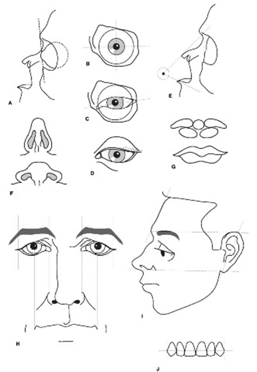
Figure 4 ‘Rules-of-thumb’ used to guide reconstruction of facial features: (A) anteroposterior placement of eyeball; (B) centering the eyeball; (C) angulation of palpebral fissure; (D) eyelids in relation to the iris; (E) finding the nose tip; (F) nostrils from below; (G) division of lip into five masses; (H) alignment between eyes, nose and mouth; (I) length of ear and its angulation; (J) lip placement over upper teeth. Refer to the text for further explanation.
Finishing off and casting
The surface of the face is smoothed and textured to resemble human skin. In order to preserve the reconstruction and make it durable for presentation, a mould is fabricated of the whole head, and a casting made in hardened plaster of Paris. Leaving the face unpainted results in an undesirable ghostly appearance. Instead, the surface of the cast is painted in flesh tones. In the absence of other clues, it is necessary to guess the color of the skin. The use of ‘add-ons’, such as facial and head hair, glasses and earrings, must be avoided at this stage. A balding head or the wrong hairstyle can confuse identity. It is preferable to have ‘add-ons’ available for placement, if requested by the identifying witness.
Discussion
It seems sensible that soft-tissue measurements be recorded on a fleshed-out living subject in the upright position, rather than on a distorted supine corpse. Also soft-tissue depths are often derived from samples of subjects ranging across age and ethnic groups. Unless samples are kept within defined and controlled parameters they are little more than a measure of neutrality. For example, average measurements on a mixed-ethnic subject iron out the differences between the primary groups and provide tissue thickness lying midway between one group and the other. In living people, this is not necessarily found to be the case. It appears the mixed individual can inherit different features from each parent. If an examination of the skull reveals an index or feature of a particular ethnic group, then the measurements and anatomical form characteristic of that group should be used for constructing the inherited feature.
There was a time when FFR was viewed with suspicion and criticism. More recently, respectability has been improved through studies that attempted to evaluate the efficacy of FFR, with varying degrees of success. Some researchers have claimed as much as a 72% success rate with the morphometric methods of reconstruction. What is meant, when a reconstruction is claimed as ‘successful’? Was it a success because it matched the deceased’s face? Was it a success because it jogged the memory of an identifying witness? Or was it a success because it played a part in excluding other suspects?
In the case illustrated in Fig. 2, the ‘success’ occurred when a policeman who saw a photograph of the reconstructed face in the newspaper remembered having seen it somewhere before. He unearthed a 17-year-old newspaper cutting of the missing person and the final identification was confirmed after photo-superimposition of the teeth. The wig had been present from the start (not advisable, as already mentioned), but the sideburns were added after the victim had been identified.
It is unfair, at this stage of its development, to expect FFR to produce an accurate positive identification. It has value in a positive sense when it stimulates direct investigative leads to the unidentified person, and in a negative sense when it eliminates those suspects whose face could not possibly ‘fit’ the unidentified skull. At all times it should be understood that facial reconstruction is still only an adjunct in the identification process. The final identification of the victim must depend on other tried and proven methods of confirmation, such as dental identification and accurate DNA analysis.
The Future
What technology and new ideas are available for research in the field of FFR? Tools such as computer tomography, magnetic resonance imaging, photo-grammetry and laser-scanning present us with masses of coordinate data on hard- and soft-tissue form. The challenging concepts of morphometrics (the fusion of geometry with biologic homology), finite elements analysis, Fourier shape analysis, neural nets and strange attractors, suggest different ways of looking at that data. The focus is swinging away from singularities (such as individual depth measurements) and is heading in the direction of continua. Even in the simplified morphometric technique (and for that matter the morphoscopic method), small but significant advances in the progress towards continuous outlines occurred though the use of drawn outlines for the lateral (and in one study, the lateral and oblique) profile of the face. The recent popularity of fuzzy logic is tempting us to discard ‘either/or’ concepts and to search for gradations of values between extremes.
The use of laser scanning to record the surface of the face, coupled with parallel processing and new Windows NT-based software, enables researchers, guided by average tissue depths, to adapt a stored face to an unknown skull and to edit and manipulate the face for a finer fit. Although speed, objectivity and the ease of circulating multiple copies of the finished result give it a certain edge over hand-sculpted efforts, there still remains the problem of using average measurements.
Sharing of visual image data between researchers in institutions and other computer stations is becoming available. A central workstation in a university computer laboratory can utilize advanced technology to produce a three-dimensional reconstruction, which can be instantly transmitted to a low-tech endpoint, for example a police station. The programs and the large amounts of data needed for such work are processed, manipulated and stored at the central terminal.
It is certainly impressive to be able to reproduce a face from masses of coordinate data. However, much of that data may well be redundant as far as an understanding of the relationship between hard and soft tissue is concerned. What should be investigated is the relationship that exists between the hard and soft tissues, which in turn suggests the formulation of predictive rules between the two. Put in another way, what is being suggested is to isolate smaller components of the skull and represent them in a form that can be statistically handled. They can then be compared with similar packages in the adjacent soft tissue and, if there is any correlation, a richer set of rules for constructing the individual features could be formulated. A sort of:’If I see this configuration on the skull, can I expect to see that type of form in the soft tissue?’ Despite the doubts of some, this may need less information than we think. As a precursor to this, clusters of similar face types could be mapped, investigated for common forms, and average measurements recorded for each cluster. Of course, right now, the human mind is more adept at noting any of these facial categories and their significance than is the computer.
Despite these forthcoming innovations, present-day clay-based forensic facial reconstructions, utilizing average soft-tissue measurements, need not be abandoned. At the very least, and for some time, it will have a role to play in areas where sophisticated technology is not freely available. And speaking of sophistication and the future, it is daring but not absurd to postulate that the rapidly developing science of DNA mapping could perhaps one day locate the genes for facial appearance.
