Dinosaur research: the scanning revolution
The steady improvement in technological resources, as well as their potential to be used to answer palaeobiological questions, has manifested in a number of distinct areas in recent years. A few of these will be examined in the following section; they are not without their limitations and pitfalls, but in some instances questions may now be asked that could not have been dreamt of 10 years ago.
One of the most anguished dilemmas faced by palaeobiologists is the desire to explore as much of any new fossil as possible, but at the same time to minimize the damage caused to the specimen by such action. The discovery of the potential for X-rays to create images on photographic film of the interior of the body has been of enormous importance to medical science. The more recent revolution in medical imaging through the development of CT (computed tomography) and MRI (magnetic resonance imaging) techniques that are linked directly to powerful data-processing computers has resulted in the ability to create three-dimensional images that allow researchers to see inside objects such as the human body or other complex structures that would only normally be possible after major exploratory surgery.
The potential to use CT scanning to see inside fossils was rapidly appreciated. One of the leaders in the field is Tim Rowe, with his team based at the University of Texas in Austin. He has managed to set up one of the finest fossil-dedicated, high-resolution CT scanning systems and, as we shall see below, has put it to some extremely interesting uses.
Investigating hadrosaurian crests
One obvious use of CT scanning can be demonstrated by referring to the extravagant range of crests seen on some hadrosaurian ornithopods. These dinosaurs were very abundant in Late Cretaceous times and have remarkably similarly shaped bodies; they only really differ in the shape of their headgear, but the reason for this difference has been a long-standing puzzle. When the first ‘hooded’ dinosaur was described in 1914, it was considered likely that these were simply interesting decorative features. However, in 1920 it was discovered that these ‘hoods’, or crests, were composed of thin sheaths of bone that enclosed tubular cavities or chambers of considerable complexity.
Theories to explain the purpose of these crests abounded from the 1920s onwards. The very earliest claimed that the crest provided an attachment area for ligaments running from the shoulders to the neck that supported the large and heavy head. From then on, ideas ranged from their use as weapons; that they carried highly developed organs of smell; that they were sexually specific (males had crests and females did not); and, the most far-sighted, that the chambers might have served as resonators, as seen in modern birds. During the 1940s, there was a preference for aquatic theories: that they formed an air-lock to prevent water flooding the lungs when these animals fed on underwater weeds.
Most of the more outlandish suggestions have been abandoned, either because physically impossible or they do not accord with the known anatomy. What has emerged is that the crests probably performed a number of interrelated functions of a mainly social/ sexual type. They probably provided a visual social recognition system for individual species; and, in addition, some elaboration of the crests undoubtedly served a sexual display purpose. A small number of hadrosaur crests were sufficiently robust to have been used either in flank or head-butting activities as part of pre-mating rituals or male-male rivalry competitions. Finally, the chambers and tubular areas associated with the crests or facial structure are thought to have functioned as resonators. Again, this presumed vocal ability (found today in birds and crocodiles) can be linked to aspects of social behaviour in these dinosaurs.
One of the greatest problems associated with the resonator theory was gaining direct access to skull material that would allow detailed reconstruction of the air passages within the crest, without breaking open prized and carefully excavated specimens. CT techniques made such internal investigations feasible. For example, some new material of the very distinctively crested hadrosaur Parasaurolophus tubicen was collected from Late Cretaceous sediments in New Mexico. The skull was reasonably complete, well preserved, and included a long, curved crest. It was CT scanned along the length of the crest, then the scans were digitally processed so that the space inside the crest, rather than the crest itself, could be imaged. The rendered version of the interior cavity revealed an extraordinary degree of complexity. Several parallel, narrow tubes looped tightly within the crest, creating the equivalent of a cluster of trombones! There is now little doubt that the crest cavities in animals like Parasaurolophus were capable of acting as resonators as part of their vocal system.
Soft tissues: hearts of stone?
In the late 1990s, a new partial skeleton of a medium-sized ornithopod was discovered in Late Cretaceous sandstones in South Dakota. Part of the skeleton was eroded away, but what remained was extraordinarily well preserved, with evidence of some of the soft tissues, such as cartilage, which are normally lost during fossilization, still visible. During initial preparation of the specimen, a large ferruginous (iron-rich) nodule was discovered in the centre of the chest. Intrigued by this structure, the researchers obtained permission to CT scan a major part of the skeleton using a large veterinary hospital scanner. The results from these scans were intriguing.
The ferruginous nodule appeared to have distinctive anatomical features, and there appeared to be associated nearby structures. The researchers interpreted these as indicating that the heart and some associated blood vessels had been preserved within the nodule. The nodule appeared to show two chambers (interpreted by the researchers as representing the original ventricles of the heart); a little above these was a curved, tube-like structure that they interpret as an aorta (one of the main arteries leaving the heart). On this basis, they went on to suggest that this showed that dinosaurs of this type had a very bird-like, fully divided heart, which supported the increasing conviction that dinosaurs were generally highly active, aerobic animals (see topic 6).
As early as 1842, and the extraordinarily prophetic speculations of Richard Owen, it had been supposed that dinosaurs, crocodiles, and birds had a relatively efficient four-chambered (i.e. fully divided) heart. On that basis, this discovery is not so startling. What is astonishing is the thought that the general shape of the soft tissues of the heart of this particular dinosaur might have been preserved through some freak circumstance of fossilization.
Soft tissue preservation is known to occur under some exceptional conditions in the fossil record; these generally comprise a mixture of very fine sediments (muds and clays) that are capable of preserving the impressions of soft tissues. Also, soft tissues, or rather their chemically replaced remnants, can be preserved by chemical precipitation, usually in the absence of oxygen. Neither of these conditions apply to the ornithopod skeleton described above. The specimen was found in coarse sandstone, and under conditions that would have been oxygen-rich, so from a simple geochemical perspective, conditions would appear to be very unlikely to preserve soft tissues of any type.
Not surprisingly, the observations made by the researchers have been challenged. Ironstone nodules are commonly reported in these deposits and are frequently found associated with dinosaur bones. The sedimentary conditions, the chemical environment in which the structures might have been preserved, and the interpretation of all the supposedly heart-like features have been contested. At present, the status of this specimen is therefore uncertain, but whatever else is claimed, if these features are simply those of an ironstone nodule, then it is extraordinary that they are so heart-like.
Fake ‘dinobirds’: forensic palaeobiology
In 1999 an article appeared in the National Geographic magazine that highlighted the similarities between dinosaurs and birds that had been revealed by the new discoveries made in Liaoning Province, China. It brought to light another new and exciting specimen that was namedArchaeoraptor, and was represented by a nearly complete skeleton that seemed as good an intermediate ‘dinobird’ as one could imagine. The animal had very bird-like wings and chest bones, yet retained rather theropod-like head, legs, and the long stiffened tail.
The specimen was initially feted by National Geographic through public events. However, the specimen soon became dogged by controversy. It had been bought by a museum based in Utah at a fossil fair in Tucson, Arizona, even though it evidently came from China. This is very unusual because the Chinese government regards all fossils of scientific value as the property of China.
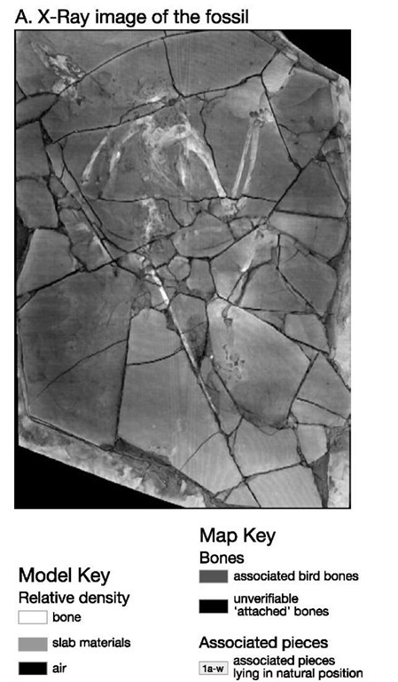
The specimen came to be regarded with suspicion by the scientific community: the front half of the body was almost too bird-like compared to the theropod-like legs and tail. The surface of limestone upon which this specimen was preserved was also unusual, it consisted of a crazy-paving-like series of small slabs held together by a lot of filler (see Figure 37). Within a relatively short period of time, it was declared to be a probable fake -possibly manufactured to order from assorted spare parts collected in Liaoning. Amid the general air of concern, the curator of the Utah museum contacted two palaeontologists who had worked on these Chinese forms, Philip Currie of the Royal Tyrell Museum, Alberta, and Xu Xing of Beijing, China; and Tim Rowe was contacted at Texas to see if he could CT scan and verify the nature of this fossil.
37. The fake Archaeoraptor on its slab of rock
By an amazing coincidence, Xu, on returning to China, located a piece of rock from Liaoning containing most of a dromaeosaur theropod. After studying this specimen, he became convinced that the tail of this fossil was the matching counterpart to the one he had recently seen on Archaeoraptor. Returning to Washington, and the office of National Geographic, Xu was able to place his recently discovered fossil against the Archaeoraptor specimen and demonstrate that the original Archaeoraptor block was without doubt a composite consisting of at least two different animals (the front half being part of a genuine bird, the back half being that of a dromaeosaur theropod).
Alerted to this, Rowe was able to study the CT scans that he made of the original Archaeoraptor slab in detail. CT cannot distinguish genuine from fraudulent fossils. However, the accuracy of the three-dimensional images of each portion of the slab allowed precise comparison of each piece of the specimen. It became clear that a partial bird fossil formed the main part of the slab, to this had been added the leg bones and feet of a theropod dinosaur. Rowe and his colleagues were able to show that only one leg bone and foot had been used. In this instance, the part and counterpart had been split apart to make a pair of legs and feet! Finally, the tail of the theropod had been added; and to complete the ‘picture’, additional pieces of paving and filler were added to create a more visually pleasing rectangular ensemble.
These dramatic revelations have had no effect whatever on the debate concerning dinosaur-bird relationships. What they do point to are some unfortunate facts. In China, where poorly paid labourers have helped to excavate some truly wondrous fossils, they have clearly developed a good knowledge of anatomy and an understanding of the sorts of creatures that scientists are looking for. These workers also realize that there is a thriving market in such fossils, which will bring them far better financial rewards if they can sell them to dealers outside China.
Dinosaur mechanics: how Allosaurus fed
Computed tomography has clearly proved to be a very valuable aid to palaeobiological investigations because it has this ability to see inside objects in an almost magical way. Some technologically innovative ways of using CT imaging have been developed by Emily Rayfield and colleagues, at the University of Cambridge. Using CT images, sophisticated computer software, and a great deal of biological and palaeobiological information, it has proved possible to investigate how dinosaurs may have functioned as living creatures.
As with the case of Tyrannosaurus, we know in very general terms that Allosaurus (Figure 31) was a predatory creature and probably fed on a range of prey living in Late Jurassic times. Sometimes tooth marks or scratches may be found on fossil bones and these can be quite literally lined up against the teeth in the jaws of an allosaur as a form of ‘proof of the guilty party. But what does such evidence tell us? The answer is: not as much as we might like. We cannot be sure if the tooth marks were left by a scavenger feeding off an already dead animal, or whether the animal that left the tooth marks was the real killer; equally, we cannot tell what style of predator an allosaur might have been: did it run down its prey after a long chase, or did it lurk and pounce? Did it have a devastating bone-crushing bite, or was it more of a cut and slasher?
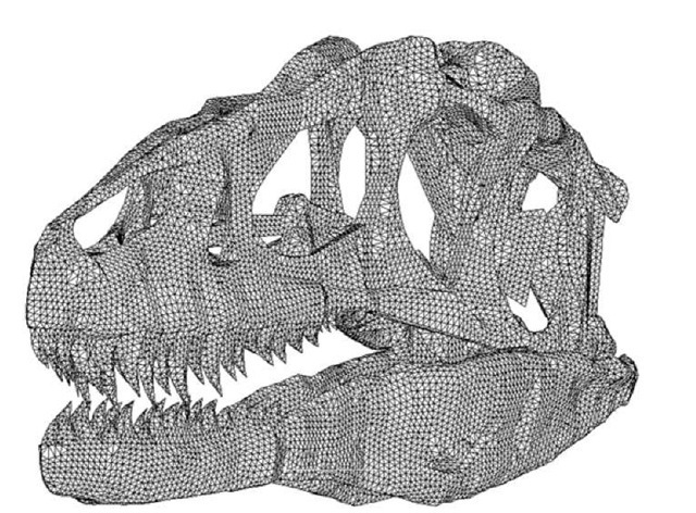
38. Finite-element modelled image of an Allosaurus skull derived from a CT scan
Rayfield was able to obtain CT scan data created from an exceptionally well-preserved skull of the Late Jurassic theropod Allosaurus. High-resolution scans of the skull were used to create a very detailed three-dimensional image of the entire skull. However, rather than simply creating a beautiful hologram-like representation of the skull, Rayfield converted the image data into a three-dimensional ‘mesh’. The mesh consisted of a series of point coordinates (rather like the coordinates on a topographic map), each point was linked to its immediate neighbours by short ‘elements’. This created what in engineering terms is known as a finite element map of the entire skull (Figure 38): nothing quite as complicated as this had ever been attempted before.
The remarkable property of this type of model is that with the appropriate computer and software it is possible to record, on the finite element map, the material properties of the skull bones, for example the strength of skull bone, of tooth enamel, or of cartilage on the joints between bones. In this way, each ‘element’ can be prompted to behave as though it were a piece of real skull, and each element is linked to its neighbours as an integrated unit, as it would be in life.
Having mapped the virtual skull of this dinosaur, it was then necessary to work out how powerful its jaw muscles were in life. Using clay, Rayfield was able to quite literally model the jaw muscles of this dinosaur. Once she had done this, she was able to calculate from their dimensions – their length, girth, and angle of attachment to the jaw bones – the amount of force that they could generate. To ensure that these calculations were as realistic as possible, two sets of force estimates were generated: one based on the view that dinosaurs like this one had a rather crocodile-like (ectotherm) physiology, the other assumed an avian/mammalian (endotherm) physiology.
Using these sets of data, it was then possible to superimpose these forces on the finite element model of the Allosaurus skull and quite literally ‘test’ how the skull would respond to maximum bite forces, and how these would be distributed within the skull. The experiments were intended to probe the construction and shape of the skull, and the way it responded to stresses associated with feeding.
What emerged was fascinating. The skull was extraordinarily strong (despite all the large holes over its surface that might be thought to have weakened it significantly). In fact, the holes proved to be an important part of the strength of the skull. When the virtual skull was tested until it began to ‘yield’ (that is to say, it was subjected to forces that were beginning to fracture its bones), it was found to be capable of withstanding up to 24 times the force that the jaw muscles could exert when they were biting as hard as ‘allosaurianly’ possible.
What became obvious from this experimentation was that the allosaur skull was hugely over-engineered. Natural selection usually provides a ‘safety factor’ in the design of most skeletal features: a sort of trade-off between the amount of energy and materials needed to build that part of the skeleton and its overall strength under normal conditions of life. That ‘safety factor’ varies, but is generally in the range of 2-5 times the forces normally experienced during normal life activities. To have the skull of Allosaurus built with a ‘safety factor’ of 24 seemed ludicrous. Re-examination of the skull, and a rethink about its potential methods of feeding, led to the following realization: the lower jaw was actually quite ‘weak’ in the way it was constructed, so the animal probably did have a genuinely weak bite, compared to its overall skull strength. This suggested that the skull was constructed to withstand very large forces (in excess of 5 tonnes) for other reasons. The most obvious was that the skull may have been used as the principal attack weapon – as a chopper. These animals may well have lunged at their prey with the jaws opened very wide, and then slammed their head downward against their prey in a devastating, slashing blow. With the weight of the body behind this movement, and the resistance of the prey animal, the skull would need to be capable of withstanding short-term, but extremely high, loads.
Once the prey had been subdued following the first attack, the jaws could then be used to bite off pieces of flesh in the conventional way, but this might reasonably have been aided by using the legs and body to assist with tugs at resistant pieces of meat, again loading the skull quite highly through forces generated by the neck, back, and leg muscles.
In this particular analysis, it has been possible to gain an idea of how feeding may have been achieved in allosaurs in ways that until a few years ago would have been unimaginable. Yet again, the interplay between new technologies and different branches of science (in this instance engineering design) can be used to probe palaeobiological problems and generate new and interesting observations.
Ancient biomolecules and tissues
I cannot finish this topic without mentioning the Jurassic Park scenario: discovering dinosaur DNA, using modern biotechnology to reconstitute that DNA, and using this to bring the dinosaur back to life.
There have been sporadic reports of finding fragments of dinosaur DNA in the scientific literature over the past decade, and then using PCR (polymerase chain reaction) biotechnology to amplify the fragments so that they can be studied more easily. Unfortunately, for those who wish to believe in the Hollywood-style scenario, absolutely none of these reports have been verified, and in truth it is exceedingly unlikely that any genuine dinosaur DNA will ever be isolated from dinosaur bone. It is simply the case that DNA is a long and complex biomolecule which degrades over time in the absence of the metabolic machinery that will maintain and repair it, as occurs in living cells. The chances of any such material surviving unaltered for over 65 million years while it is buried in the ground (and subject there to all the contamination risks presented by micro-organisms and other biological and chemical sources, and ground water) are effectively zero.
All reports of dino-DNA to date have proved to be records of contaminants. In fact the only reliable fossil DNA that has been identified is far more recent, and even these discoveries have been made possible because of unusual preservational conditions. For example, brown bear fossils whose remains are dated back to about 60,000 years have yielded short strings of mitochondrial DNA – but these fossils had been frozen in permafrost since the animals died, providing the best chance of reducing the rate of degradation of these molecules. Dinosaur remains are of course 1,000 times more ancient than those of arctic brown bears. Although it might be possible to identify some dinosaur-like genes in the DNA of living birds, regenerating a dinosaur is beyond the bounds of science.
One final, but extremely interesting, set of observations concerns the analysis of the appearance and chemical composition of the interior of some tyrannosaur bones from Montana. Mary Schweitzer and colleagues from North Carolina State University were given access to some remarkably well preserved T. rex bones collected by Jack Horner (the real-life model for ‘Dr Alan Grant’ in the film Jurassic Park). Detailed examination of the skeletal remains suggested that there had been minimal alteration of the internal structure of the long bones; indeed, so unaltered were they that the individual bones of the tyrannosaur had a density that was consistent with that of modern bones that had simply been left to dry.
Schweitzer was looking for ancient biomolecules, or at least the remnant chemical signals that they might have left behind. Having extracted material from the interior of the bones, this was powdered and subjected to a broad range of physical, chemical, and biological analyses. The idea behind this approach was not only to have the best chance of ‘catching’ some trace, but also to have a range of semi-independent support for the signal, if it emerged. The burden really is upon the researcher to find some positive proof of the presence of such biomolecules; the time elapsed since death and burial, and the overwhelming probability that any remnant of such molecules has been completely destroyed or flushed away, seem to be overwhelming. Nuclear magnetic resonance and electron spin resonance revealed the presence of molecular residues resembling haemoglobin (the primary chemical constituent of red blood cells); spectroscopic analysis and HPLC (high performance liquid chromatography) generated data that was also consistent with the presence of remnants of the haeme structure. Finally, the dinosaur bone tissues were flushed with solvents to extract any remaining protein fragments; this extract was then injected into laboratory rats to see if it would raise an immune response – and it did! The antiserum created by the rats reacted positively with purified avian and mammalian haemoglobins. From this set of analyses, it seems very probable that chemical remnants of dinosaurian haemoglobin compounds were preserved in these T. rex tissues.
Even more tantalizingly, when thin sections of portions of bone were examined microscopically, small, rounded microstructures could be identified in the vascular channels (blood vessels) within the bone. These microstructures were analysed and found to be notably iron-rich compared to the surrounding tissues (iron being a principal constituent of the haeme molecule). Also the size and general appearance was remarkably reminiscent of avian nucleated blood cells. Although these structures are not actual blood cells, they certainly seem to be the chemically altered ‘ghosts’ of the originals. Quite how these structures have survived in this state for 65 Ma is a considerable puzzle.
Schweitzer and her co-workers have also been able to identify (using immunological techniques similar to the one mentioned above) biomolecular remnants of the ‘tough’ proteins known as collagen (a major constituent of natural bone, as well as ligaments and tendons) and keratin (the material that forms scales, feathers, hair, and claws).
Although these results have been treated with considerable scepticism by the research community at large – and rightly so, for the reasons elaborated above – nevertheless, the range of scientific methodologies employed to support their conclusions, and the exemplary caution with which these observations were announced, represent a model of clarity and application of scientific methodologies in this field of palaeobiology.


