Mesial Aspect
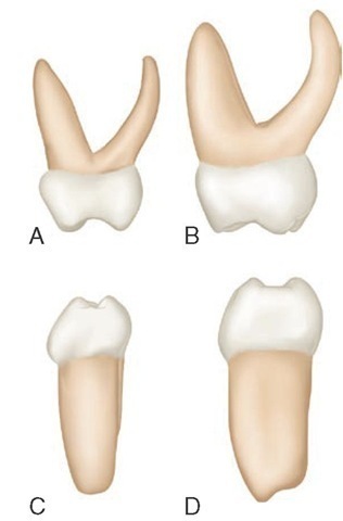
From the mesial aspect, the dimension at the cervical third is greater than the dimension at the occlusal third (Figure 3-23, A). This is true of all molar forms, but it is more pronounced on primary than on permanent teeth. The mesio-lingual cusp is longer and sharper than the mesiobuccal cusp. A pronounced convexity is evident on the buccal outline of the cervical third. This convexity is an outstanding characteristic of this tooth. It actually gives the impression of overdevelopment in this area when comparisons are made with any other tooth, primary or permanent, with the mandibular first primary molar being a close contender. The cervical line mesially shows some curvature in the direction of the occlusal surface.
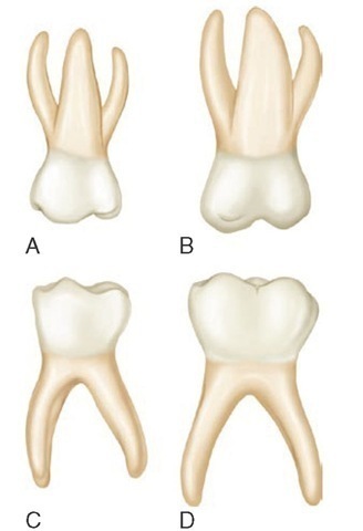
Figure 3-22 Primary right molars, lingual aspect. A, Maxillary first molar. B, Maxillary second molar. C, Mandibular first molar. D, Mandibular second molar.
The mesiobuccal and lingual roots are visible only when looking at the mesial side of this tooth from a point directly opposite the contact area. The distobuccal root is hidden behind the mesiobuccal root. The lingual root from this aspect looks long and slender and extends lingually to a marked degree. It curves sharply in a buccal direction above the middle third.
Figure 3-23 Primary right molars, mesial aspect. A, Maxillary first molar. B, Maxillary second molar. C, Mandibular first molar. D, Mandibular second molar.
Distal Aspect
From the distal aspect, the crown is narrower distally than mesially; it tapers markedly toward the distal end (Figure 3-24, A). The distobuccal cusp is long and sharp, and the distolingual cusp is poorly developed. The prominent bulge seen from the mesial aspect at the cervical third does not continue distally. The cervical line may curve occlusally, or it may extend straight across from the buccal surface to the lingual surface. All three roots may be seen from this angle, but the distobuccal root is superimposed on the mesiobuccal root so that only the buccal surface and the apex of the latter may be seen. The point of bifurcation of the distobuccal root and the lingual root is near the CEJ and, as described earlier, is typical.
Occlusal Aspect
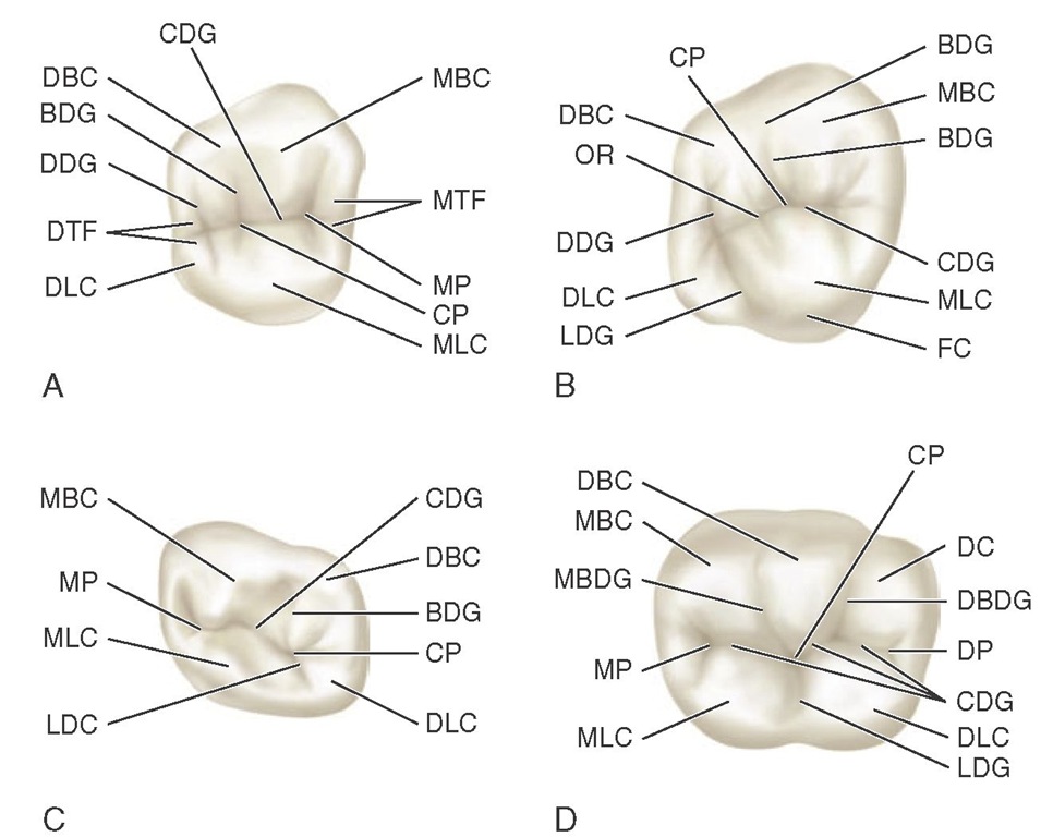
The calibration of the distance between the mesiobuccal line angle and the distobuccal line angle is definitely greater than the calibration between the mesiolingual line angle and the distolingual line angle (see Figure 3-24, A). Therefore the crown outline converges lingually. Also, the calibration from the mesiobuccal line angle to the mesiolingual line angle is definitely greater than that found at the distal line angles. Therefore the crown also converges distally. Nevertheless, these convergences are not reflected entirely in the working occlusal surface because it is more nearly rectangular, with the shortest sides of the rectangle represented by the marginal ridges. The occlusal surface has a central fossa. A mesial triangular fossa is just inside the mesial marginal ridge, with a mesial pit in this fossa and a sulcus with its central groove connecting the two fossae. A well-defined buccal developmental groove divides the mesiobuccal cusp and the distobuccal cusp occlusally. Supplemental grooves radiate from the pit in the mesial triangular fossa as follows: one buccally, one lingually, and one toward the marginal ridge, with the last sometimes extending over the marginal ridge mesially.
Figure 3-24 A, Maxillary first molar. MBC, Mesiobuccal cusp; MTF, mesial triangular fossa; MP, mesial pit; CP, central pit; MLC, mesiolingual cusp; DLC, distolingual cusp; DTF, distal triangular fossa; DDG, distal developmental groove; BDG, buccal developmental groove; DBC, distobuccal cusp; CDG, central developmental groove. B, Maxillary second molar. BDG, Buccal developmental groove; MBC, mesiobuccal cusp; CDG, central developmental groove; MLC, mesiolingual cusp; FC, fifth cusp; LDG, lingual developmental groove; DLC, distolingual cusp; DDG, distal developmental groove; OR, oblique ridge; DBC, distobuccal cusp; CP, central pit. C, Mandibular first molar. CDG, Central developmental groove; DBC, distobuccal cusp; BDG, buccal developmental groove; CP, central pit; DLC, distolingual cusp; LDC, lingual developmental groove; MLC, mesiolingual cusp; MP, mesial pit; MBC, mesiobuccal cusp. D, Mandibular second molar. DBC, Distobuccal cusp; CP, central pit; DC, distal cusp; DBDG, distobuccal developmental groove; DP, distal pit; CDG, central developmental groove; DLC, distolingual cusp; LDG, lingual developmental groove; MLC, mesiolingual cusp; MP, mesial pit; MBDG, mesiobuccal developmental groove; MBC, mesiobuccal cusp.
Sometimes the primary maxillary first molar has a well-defined triangular ridge connecting the mesiolingual cusp with the distobuccal cusp. When well developed, it is called the oblique ridge. In some of these teeth, the ridge is indefinite, and the central developmental groove extends from the mesial pit to the distal developmental groove. This disto-occlusal groove is always seen and may or may not extend through to the lingual surface, outlining a distolingual cusp. The distal marginal ridge is thin and poorly developed in comparison with the mesial marginal ridge.
Summary of the occlusal aspect of the maxillary first primary molar
The form of the maxillary first primary molar varies from that of any tooth in the permanent dentition. Although no premolars are in the primary set, in some respects the crown of this primary molar resembles a permanent maxillary premolar. Nevertheless, the divisions of the occlu-sal surface and the root form with its efficient anchorage make it a molar, both in type and function. Figure 3-25 presents all the aspects of the primary maxillary first molars.
MAXILLARY SECOND MOLAR
Buccal Aspect
The primary maxillary second molar has characteristics resembling those of the permanent maxillary first molar, but it is smaller (see Figure 3-21, B). The buccal view of this tooth shows two well-defined buccal cusps with a buccal developmental groove between them (Figure 3-26, 1). In line with all primary molars, the crown is narrow at the cervix in comparison with its mesiodistal measurement at the contact areas. This crown is much larger than that of the first primary molar. Although from this aspect the roots appear slender, they are much longer and heavier than those that are a part of the maxillary first molar. The point of bifurcation between the buccal roots is close to the cervical line of the crown. The two buccal cusps are more nearly equal in size and development than those of the primary maxillary first molar.
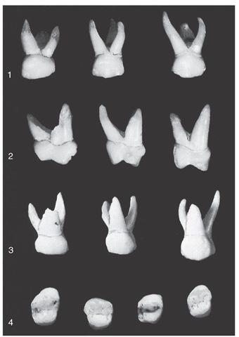
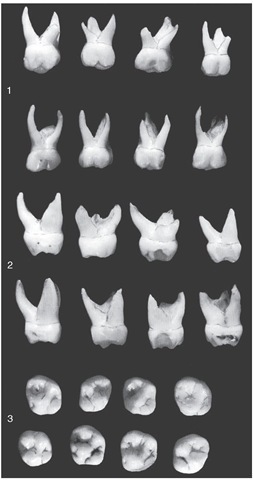
Figure 3-25 Primary maxillary first molars. 1, Buccal aspect. Note the flare of roots. 2, Mesial aspect. The cervical ridge on the buccal surface is curved to the extreme. Also note the flat or concave buccal surface above this bulge as it approaches the occlusal surface. 3, Lingual aspect. 4, Occlusal aspect. This aspect emphasizes the extensive width of the mesial portion of primary first molars. The four specimens show size differentials even in deciduous teeth (see Figure 3-24).
Lingual Aspect
Lingually, the crown shows the following three cusps:
(1) the mesiolingual cusp, which is large and well developed;
(2) the distolingual cusp, which is well developed (more so than that of the primary first molar); and (3) a third supplemental cusp, which is apical to the mesiolingual cusp and sometimes called the tubercle of Carabelli, or the fifth cusp (see Figure 3-22, B). This cusp is poorly developed and merely acts as a buttress or supplement to the bulk of the mesiolingual cusp. If the tubercle of Carabelli seems to be missing, some traces of developmental lines or "dimples" remain (see Figure 3-26, 3). A well-defined developmental groove separates the mesiolingual cusp from the distolingual cusp and connects with the developmental groove, which outlines the fifth cusp.
All three roots are visible from this aspect; the lingual root is large and thick in comparison with the other two roots. It is approximately the same length as the mesiobuccal root. If it should differ, it will be on the short side.
Figure 3-26 Primary maxillary second molars. 1, Buccal aspect. 2, Mesial aspect. 3, Occlusal aspect.
Mesial Aspect
From the mesial aspect, the crown has a typical molar outline that resembles that of the permanent molars very much (see Figures 3-23, B and 3-26, 2). The crown appears short because of its width buccolingually in comparison with its length. The crown of this tooth is usually only about 0.5 mm longer than the crown of the first deciduous molar, but the buccolingual measurement is 1.5 to 2 mm greater. In addition, the roots are 1.5 to 2 mm longer. The mesiolingual cusp of the crown with its supplementary fifth cusp appears large in comparison with the mesiobuccal cusp. The mesiobuccal cusp from this angle is relatively short and sharp. Little curvature to the cervical line is evident. Usually, it is almost straight across from buccal surface to lingual surface.
The mesiobuccal root from this aspect is broad and flat. The lingual root has somewhat the same curvature as the lingual root of the maxillary first deciduous molar.
The mesiobuccal root extends lingually far out beyond the crown outline. The point of bifurcation between the mesiobuccal root and the lingual root is 2 or 3 mm apical to the cervical line of the crown; this differs in depth on the root trunk from comparisons of this area in the recent discussion of primary molars. The mesiobuccal root presents itself as being quite wide from the mesial aspect. It measures approximately two thirds the width of the root trunk, which leaves one third for the lingual root. The mesiolingual cusp is directly below their bifurcation. Although from this aspect the curvature is strong lingually at the cervical portion, as on most deciduous teeth the crest of curvature buccally at the cervical third is nominal and resembles the curvature found at this point on the permanent maxillary first molar. In this, it differs entirely from the prominent curvature found on the primary maxillary first molars at the cervical third buccally.
Distal Aspect
From the distal aspect, it is apparent that the distal calibration of the crown is less than the mesial measurement, but the variation is found on the crown of the deciduous maxillary first molar. From both the distal and the mesial aspects, the outline of the crown lingually creates a smooth, rounded line, whereas a line describing the buccal surface is almost straight from the crest of curvature to the tip of the buccal cusp. The distobuccal cusp and the distolingual cusp are about the same in length. The cervical line is approximately straight, as was found mesially.
All three roots are seen from this aspect, although only a part of the outline of the mesiobuccal root may be seen, since the distobuccal root is superimposed over it. The dis-tobuccal root is shorter and narrower than the other roots. The point of bifurcation between the distobuccal root and the lingual root is more apical in location than any of the other points of bifurcation. The point of bifurcation between these two roots on the distal is more nearly centered above the crown than that on the mesial between the mesiobuccal and lingual roots.
