Maxillary Canine
Labial Aspect
Except for the root form, the labial aspect of the maxillary canine does not resemble either the central or the lateral incisor (Figure 3-16; see also Figure 3-11, C). The crown is more constricted at the cervix in relation to its mesiodistal width, and the mesial and distal surfaces are more convex. Instead of an incisal edge that is relatively straight, it has a long, well-developed, sharp cusp.
Compared with that of the permanent maxillary canine, the cusp on the primary canine is much longer and sharper, and the crest of contour mesially is not as far down toward the incisal portion. A line drawn through the contact areas of the deciduous canine would bisect a line drawn from the cervix to the tip of the cusp. In the permanent canine, the contact areas are not at the same level. When the cusp is intact, the mesial slope of the cusp is longer than the distal slope. The root of the primary canine is long, slender, and tapering and is more than twice the crown length.
Lingual Aspect
The lingual aspect shows pronounced enamel ridges that merge with each other (see Figure 3-12, C). They are the cingulum, mesial and distal marginal ridges, and incisal cusp ridges, besides a tubercle at the cusp tip, which is a continuation of the lingual ridge connecting the cingulum and the cusp tip. This lingual ridge divides the lingual surface into shallow mesiolingual and distolingual fossae.
The root of this tooth tapers lingually. It is usually inclined distally also above the middle third (see Figures 3-11, C and 3-13, C).
Mesial Aspect
From the mesial aspect, the outline form is similar to that of the lateral and central incisors (see Figures 3-13, C and 3-16, 2). However, a difference in proportion is evident. The measurement labiolingually at the cervical third is much greater. This increase in crown dimension, in conjunction with the root width and length, permits resistance against forces the tooth must withstand during function. The function of this tooth is to punch, tear, and apprehend food material.
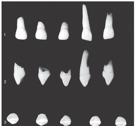
Figure 3-15 Primary maxillary lateral incisors (second incisors). 1, Labial aspect. 2, Mesial aspect. 3, Incisal aspect.
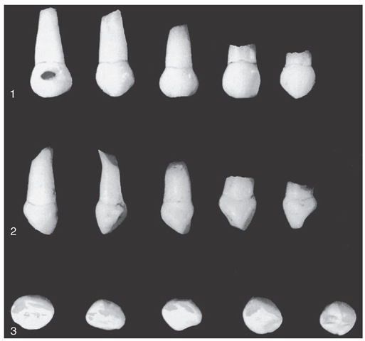
Figure 3-16 Primary mandibular central incisors. 1, Labial aspect. 2, Mesial aspect. 3, Incisal aspect.
Distal Aspect
The distal outline of this tooth is the reverse of the mesial aspect. No outstanding differences may be noted except that the curvature of the cervical line toward the cusp ridge is less than on the mesial surface.
Incisal Aspect
From the incisal aspect, we observe that the crown is essentially diamond-shaped (see Figures 3-14, C and 3-16, 3). The angles that are found at the contact areas mesially and distally; the cingulum on the lingual surface; and the cervical third, or enamel ridge, on the labial surface are more pronounced and less rounded in effect than those found on the permanent canines. The tip of the cusp is distal to the center of the crown, and the mesial cusp slope is longer than the distal cusp slope. This allows for intercuspation with the lower, or mandibular, canine, which has its longest slope distally (see Figure 3-11).
MANDIBULAR CENTRAL INCISOR
Labial Aspect
The labial aspect of this crown has a flat face with no developmental grooves (Figure 3-17; see Figure 3-11, D). The mesial and distal sides of the crown are tapered evenly from the contact areas, with the measurement being less at the cervix. This crown is wide in proportion to its length in comparison with that of its permanent successor. The heavy look at the root trunk makes this small tooth resemble the permanent maxillary lateral incisor.
The root of the primary central incisor is long and evenly tapered down to the apex, which is pointed. The root is almost twice the length of the crown (see Figure 3-11, D).
Lingual Aspect
On the lingual surface of the crown, the marginal ridges and the cingulum may be located easily (see Figure 3-12, D). The lingual surface of the crown at the middle third and the incisal third may have a flattened surface level with the marginal ridges, or it may present a slight concavity, called the lingual fossa. The lingual portion of the crown and root converges so that it is more narrow toward the lingual and not the labial surface.
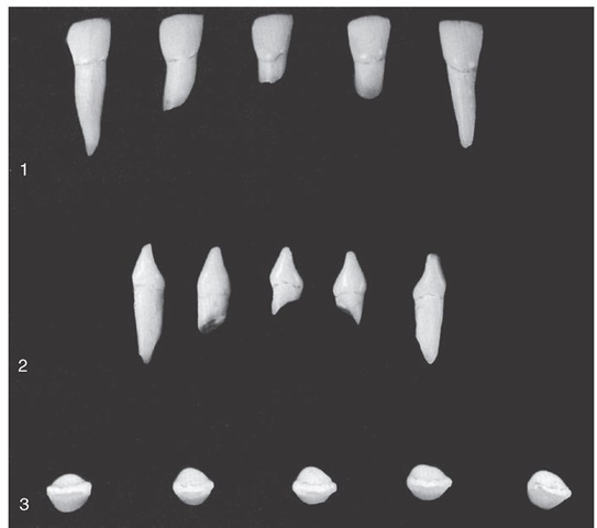
Figure 3-17 Primary mandibular central incisors. 1, Labial aspect. 2, Mesial aspect. 3, Incisal aspect.
Mesial Aspect
The mesial aspect shows the typical outline of an incisor tooth, even though the measurements are small (see Figures 3-13, D and 3-17, 2). The incisal ridge is centered over the center of the root and between the crest of curvature of the crown, labially, and lingually. The convexity of the cervical contours labially and lingually at the cervical third is just as pronounced as in any of the other primary incisors and more pronounced by far than the prominences found at the same locations on a permanent mandibular central incisor. As mentioned before, these cervical bulges are important.
Although this tooth is small, its labiolingual measurement is only about a millimeter less than that of the primary maxillary central incisor. The primary incisors seem to be built for strenuous service.
The mesial surface of the root is nearly flat and evenly tapered; the apex presents a more blunt appearance than is found with the lingual or labial aspects.
Distal Aspect
The outline of this tooth from the distal aspect is the reverse of that found from the mesial aspect. Little difference can be noted between these aspects, except that the cervical line of the crown is less curved toward the incisal ridge than on the mesial surface. Often, a developmental depression is evident on the distal side of the root.
Incisal Aspect
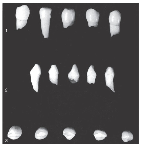
The incisal ridge is straight and bisects the crown labiolin-gually. The outline of the crown from the incisal aspect emphasizes the crests of contour at the cervical third labially and lingually (see Figures 3-14, D and 3-17, 3). A definite taper is evident toward the cingulum on the lingual side.
The labial surface from this view presents a flat surface that is slightly convex, whereas the lingual surface presents a flattened surface that is slightly concave.
MANDIBULAR LATERAL INCISOR
The fundamental outlines of the primary mandibular lateral incisor (Figure 3-18) are similar to those of the primary central incisor. These two teeth supplement each other in function. The lateral incisor is somewhat larger in all measurements except labiolingually, where the two teeth are practically identical. The cingulum of the lateral incisor may be a little more generous than that of the central incisor. The lingual surface of the crown between the marginal ridges may be more concave. In addition, a tendency exists for the incisal ridge to slope downward distally. This design lowers the distal contact area apically, so that proper contact may be made with the mesial surface of the primary mandibular canine (see Figures 3-11, E; 3-12, E; 3-13, E; and 3-14, E).
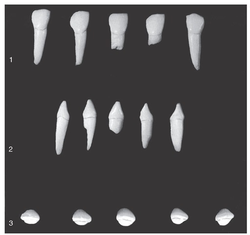
Figure 3-18 Primary mandibular lateral incisors. 1, Labial aspect. 2, Mesial aspect. 3, Incisal aspect.
Mandibular Canibe
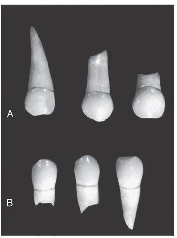
Little difference in functional form is evident between the mandibular canine and the maxillary canine. The difference is mainly in the dimensions. The crown is perhaps 0.5 mm shorter, and the root is at least 2 mm shorter; the mesiodistal measurement of the mandibular canine at the root trunk is greater when compared with its mesiodistal measurement at the contact areas than is that of the maxillary canine (Figure 3-19). It is "thicker" accordingly at the "neck" of the tooth. The outstanding variation in size between the two deciduous canines is shown by the labiolingual calibration. The deciduous maxillary canine is much larger labiolingually (see Figure 3-13).
The cervical ridges labially and lingually are not quite as pronounced as those found on the maxillary canine. The greatest variation in outline form when one compares the two teeth is seen from the labial and lingual aspects; the distal cusp slope is longer than the mesial slope. The opposite arrangement is true of the maxillary canine. This makes for proper intercuspation of these teeth during mastication.
Figure 3-19 A comparison of primary canines, both in the size and shape of the crowns. Two of them have their roots intact and show no dissolution. A, Maxillary canines. B, Mandibular canines. Compare Figures 3-16 and 3-20.
Figure 3-20 Primary mandibular central incisors. 1, Labial aspect. 2, Mesial aspect. 3, Incisal aspect.
MAXILLARY FIRST MOLAR
Buccal Aspect
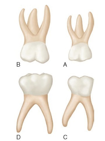
The widest measurement of the crown of the maxillary first molar is at the contact areas mesially and distally (Figure 3-21, A). From these points, the crown converges toward the cervix, with the measurement at the cervix being fully 2 mm less than the measurement at the contact areas. This dimensional arrangement furnishes a narrower look to the cervical portion of the crown and root of the primary maxillary first molar than that of the same portion of the permanent maxillary first molar. The occlusal line is slightly scalloped but with no definite cusp form. The buccal surface is smooth, and little evidence of developmental grooves is noted. It is from this aspect that one may judge the relative size of the primary maxillary first molar when it is compared with the second molar. It is much smaller in all measurements than the second molar. Its relative shape and size suggest that it was designed to be the "premolar section" of the primary dentition. In function it acts as a compromise between the size and shape of the anterior primary teeth and the molar area, this area being held temporarily by the larger primary second molar. At age 6, the large first permanent molar is expected to take its place distal to the second primary molar, which will complete a more extensive molar area for masticating efficiency.
The roots of the maxillary first molar are slender and long, and they spread widely. All three roots may be seen from this aspect. The distal root is considerably shorter than the mesial one. The bifurcation of the roots begins almost immediately at the site of the cervical line (CEJ). Actually, this arrangement is in effect for the entire root trunk, which includes a trifurcation, and this is a characteristic of all primary molars, whether maxillary or mandibular. Permanent molars do not possess this characteristic. The root trunk on permanent molars is much heavier, with a greater distance between the cervical lines to the points of bifurcations (see Figure 11-8).
Figure 3-21 Primary right molars, buccal aspect. A, Maxillary first molar. B, Maxillary second molar. C, Mandibular first molar. D, Mandibular second molar.
Lingual Aspect
The general outline of the lingual aspect of the crown is similar to that of the buccal aspect (Figure 3-22, A). The crown converges considerably in a lingual direction, which makes the lingual portion calibrate less mesiodistally than the buccal portion.
The mesiolingual cusp is the most prominent cusp on this tooth. It is the longest and sharpest cusp. The distolin-gual cusp is poorly defined; it is small and rounded when it exists at all. From the lingual aspect, the distobuccal cusp may be seen, since it is longer and better developed than the distolingual cusp. A type of primary maxillary first molar that is not uncommon and presents as one large lingual cusp with no developmental groove in evidence lingually is a three-cusped molar (see Figure 3-25, 4, second from left).
All three roots also may be seen from this aspect. The lingual root is larger than the others.
