The maxillary premolars number four: two in the right maxilla and two in the left maxilla. They are posterior to the canines and immediately anterior to the molars.
The premolars are so named because they are anterior to the molars in the permanent dentition. In zoology, the premolars are those teeth that succeed the deciduous molars regardless of the number to be succeeded. The term bicuspid, which is widely used to describe human teeth, presupposes two cusps, a supposition that makes the term misleading, because mandibular premolars in the human subject may show a variation in the number of cusps from one to three. Among carnivores, in the study of comparative dental anatomy, premolar forms differ so greatly that a more descriptive single term than premolar is out of the question. The term premolar is used widely in dental anatomy, human and comparative; therefore it will be used here. However, its use here does not suggest that the term bicuspid should not be used when appropriate.
The maxillary premolars are developed from the same number of lobes as anterior teeth—four. The primary difference in development is the well-formed lingual cusp, developed from the lingual lobe, which is represented by the cingulum development on incisors and canines. The middle buccal lobe on the premolars, corresponding to the middle labial lobe of the canines, remains highly developed, with the maxillary premolars resembling the canines when viewed from the buccal aspect. The buccal cusp of the maxillary first premolar, especially, is long and sharp, assisting the canine as a prehensile or tearing tooth. The mandibular first premolar assists the mandibular canine in the same manner.
The second premolars, both maxillary and mandibular, have cusps less sharp than the others, and their cusps articulate with opposing teeth when the jaws are brought together; this makes them more efficient as grinding teeth, and they function much like the molars, but to a lesser degree.
The maxillary premolar crowns are shorter than those of the maxillary canines, and the roots are also shorter. The root lengths equal those of the molars. The crowns are a little longer than those of the molars.
Because of the cusp development buccally and lingually, the marginal ridges are in a more horizontal plane and are considered part of the occlusal surface of the crown rather than part of the lingual surface, as in the case of incisors and canines.
When premolars have two roots, one is placed buccally and one lingually.
Maxillary First Premolar
Figures 9-1 through 9-16 illustrate the maxillary first premolar from all aspects. The maxillary first premolar has two cusps, a buccal and a lingual, each being sharply defined. The buccal cusp is usually about 1 mm longer than the lingual cusp. The crown is angular, and the buccal line angles are prominent.
The crown is shorter than that of the canine by 1.5 to 2 mm on the average (Table 9-1). Although this tooth resembles the canine from the buccal aspect, it differs in that the contact areas mesially and distally are at about the same level. The root is shorter. If the buccal cusp form has not been changed by wear, the mesial slope of the cusp is longer than the distal slope. The opposite arrangement is true of the maxillary canine. Generally, the first premolar is not as wide in a mesiodistal direction as the canine.
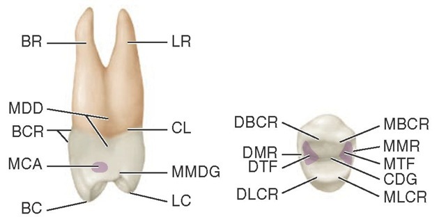
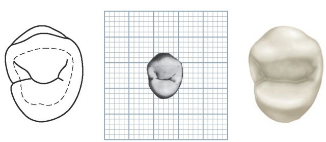
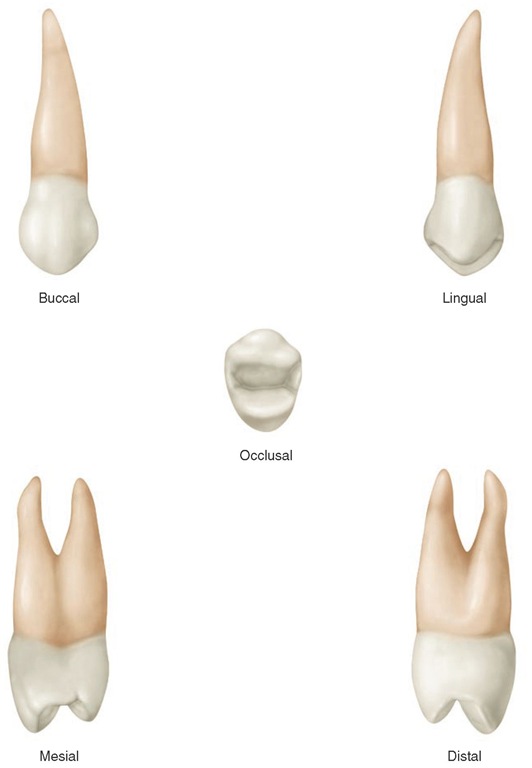
FIGURE 9-1 Maxillary right first premolar, mesial and occlusal aspects. LR, Lingual root; CL, cervical line; MMDG, mesial marginal developmental groove; LC, lingual cusp; BC, buccal cusp; MCA, mesial contact area; BCR, buccal cervical ridge; MDD, mesial developmental depression; BR, buccal root; MBCR, mesiobuccal cusp ridge; MMR, mesial marginal ridge; MTF, mesial triangular fossa (shaded area); CDG, central developmental groove; MLCR, mesiolingual cusp ridge; DLCR, distolingual cusp ridge; DTF, distal triangular fossa; DMR, distal marginal ridge; DBCR, distobuccal cusp ridge.
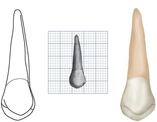
FIGURE 9-2 Maxillary left first premolar, buccal aspect. (Grid = 1 sq mm.)
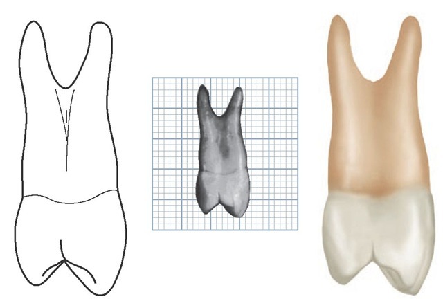
FIGURE 9-3 Maxillary left first premolar, lingual aspect. (Grid = 1 sq mm.)
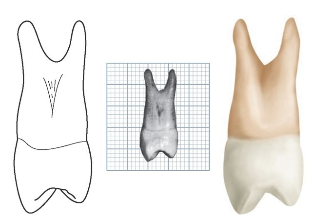
FIGURE 9-4 Maxillary left first premolar, mesial aspect. (Grid = 1 sq mm.)
FIGURE 9-5 Maxillary left first premolar, distal aspect. (Grid = 1 sq mm.)
FIGURE 9-6 Maxillary left first premolar, occlusal aspect. (Grid = 1 sq mm.)
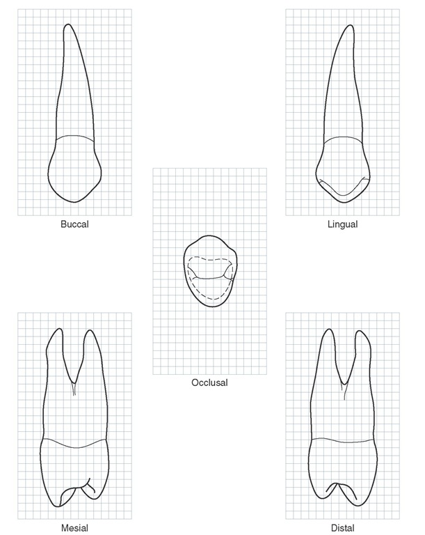
Figure 9-7 Maxillary right first premolar. Graph outlines of five aspects are shown. (Grid = 1 sq mm.)
Table 9-1 Maxillary First Premolar
Figure 9-8 Maxillary right first premolar.
Most maxillary first premolars have two roots (see Figure 9-10) and two pulp canals. When only one root is present, two pulp canals are usually found anyway.
The maxillary first premolar has some characteristics common to all posterior teeth. Briefly, those characteristics that differentiate posterior teeth from anterior teeth are as follows:
1. Greater relative faciolingual measurement compared with the mesiodistal measurement
2. Broader contact areas
3. Contact areas more nearly at the same level
4. Less curvature of the cervical line mesially and distally
5. Shorter crown, cervico-occlusally than that of anterior teeth
Detailed Description of the Maxillary First Premolar From All Aspects
Buccal Aspect
From the buccal aspect, the crown is roughly trapezoidal (see Figure 4-16, C). The crown exhibits little curvature at the cervical line. The crest of curvature of the cervical line buccally is near the center of the root buccally (see Figure 9-2 and Figures 9-7 through 9-9).
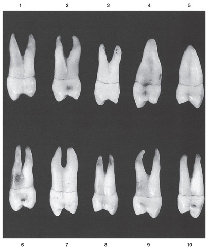
Figure 9-9 Maxillary first premolar, buccal aspect. Ten typical specimens are shown.
The mesial outline of the crown is slightly concave from the cervical line to the mesial contact area. The contact area is represented by a relatively broad curvature, the crest of which lies immediately occlusal to the halfway point from the cervical line to the tip of the buccal cusp.
The mesial slope of the buccal cusp is rather straight and longer than the distal slope, which is shorter and more curved. This arrangement places the tip of the buccal cusp distal to a line bisecting the buccal surface of the crown. The mesial slope of the buccal cusp is sometimes notched; in other instances, a concave outline is noted at this point (see Figure 9-9, 9, and 10).
The distal outline of the crown below the cervical line is straighter than that of the mesial, although it may be somewhat concave also. The distal contact area is represented by a broader curvature than is found mesially, and the crest of curvature of the contact area tends to be a little more occlusal when the tooth is posed with its long axis vertical. Even so, the contact areas are more nearly level with each other than those found on anterior teeth.
The width of the crown of the maxillary first premolar mesiodistally is about 2 mm less at the cervix than at its width at the points of its greatest mesiodistal measurement.
The buccal cusp is long, coming to a pointed tip and resembling the canine in this respect, although contact areas in this tooth are near the same level.
The buccal surface of the crown is convex, showing strong development of the middle buccal lobe. The continuous ridge from cusp tip to cervical margin on the buccal surface of the crown is called the buccal ridge.
FIGURE 9-10 Maxillary first premolar, mesial aspect. Ten typical specimens are shown.
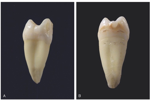
FIGURE 9-11 A, Mesial view of a maxillary right first premolar demonstrating single root and mesial crown concavity. B, Mesial view of a maxillary right second premolar demonstrating single root and no mesial crown concavity.
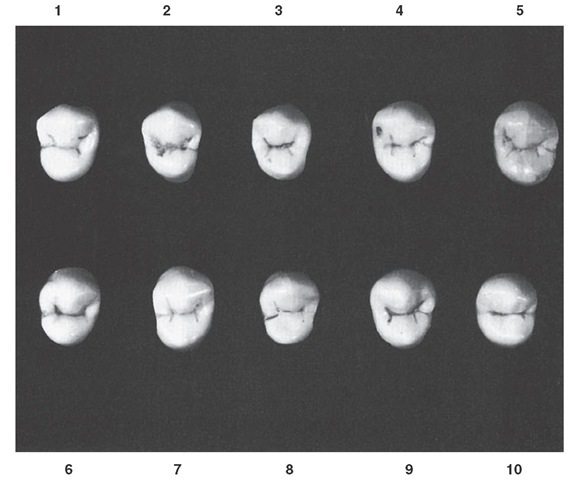
Figure 9-12 Maxillary first premolar, occlusal aspect. Ten typical specimens are shown.
Mesial and distal to the buccal ridge, at or occlusal to the middle third, developmental depressions are usually seen that serve as demarcations between the middle buccal lobe and the mesiobuccal and distobuccal lobes. Although the latter lobes show less development, they are nevertheless prominent and serve to emphasize strong mesiobuccal and distobuccal line angles on the crown.
The roots are 3 or 4 mm shorter than those of the maxillary canine, although the outline of the buccal portion of the root form bears a close resemblance.
