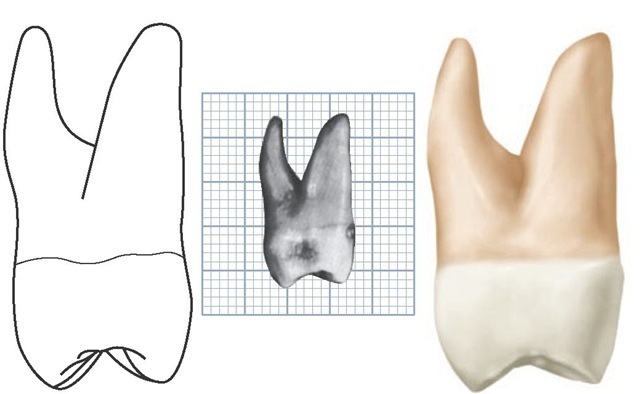
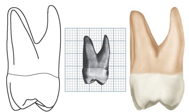
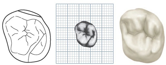
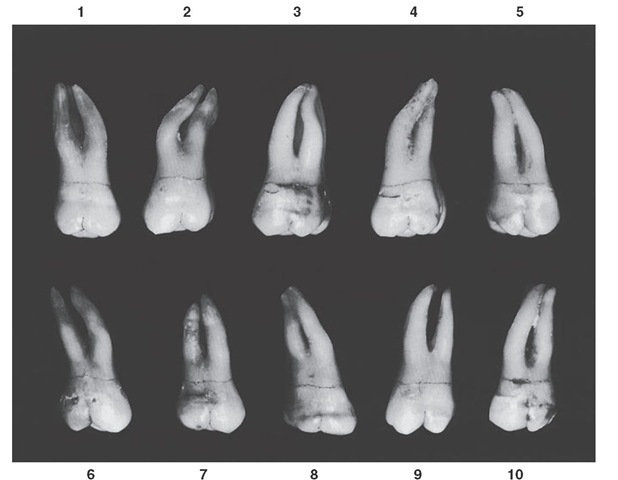
Distal Aspect
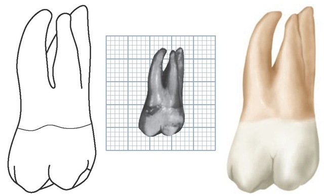
The gross outline of the distal aspect is similar to that of the mesial aspect (see Figures 11-9, 11-10, 11-13, and 11-14). Certain variations must be noted when the tooth is viewed from the distal aspect.
Because of the tendency of the crown to taper distally on the buccal surface, most of the buccal surface of the crown may be seen in perspective from the distal aspect. This is because the buccolingual measurement of the crown mesially is greater than the same measurement distally. All of the decrease in measurement distally is a result of the slant of the buccal side of the crown.
The distal marginal ridge dips sharply in a cervical direction, exposing triangular ridges on the distal portion of the occlusal surface of the crown.
The cervical line is almost straight across from buccal to lingual. Occasionally it curves apically 0.5 mm or so.
The distal surface of the crown is generally convex, with a smoothly rounded surface except for a small area near the distobuccal root at the cervical third. This concavity continues on to the distal surface of the distobuccal root, from the cervical line to the area of the root that is on a level with bifurcation separating the distobuccal and lingual roots.
The distobuccal root is narrower at its base than either of the others. An outline of this root, when the tooth is viewed from the distal aspect, starts buccally at a point immediately above the distobuccal cusp, follows a concave path inward for a short distance, then turns outward in a buccal direction, completing a graceful convex arc from the concavity to the rounded apex. This line lies entirely within the confines of the outline of the mesiobuccal root. The lingual outline of the root from the apex to the bifurcation is slightly concave. No concavity is evident between the bifurcation of the roots and the cervical line. However, the surface at this point on the root trunk has a tendency toward convexity.
The bifurcation here is more apical than either of the other two areas on this tooth. The area from cervical line to bifurcation is 5 mm or more in extent.
Occlusal Aspect
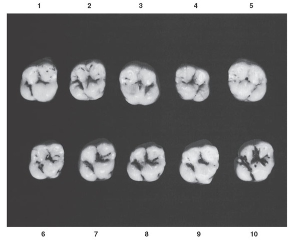
From the occlusal aspect, the maxillary first molar is somewhat rhomboidal. An outline following the four major cusp ridges and the marginal ridges is especially so (see Figures 11-1, 11-2, 11-12, 11-13, 11-14, and 11-17).
A measurement of the crown buccolingually and mesial to the buccal and lingual grooves is greater than the measurement on that portion of the crown which is distal to these developmental grooves. Also, a measurement of the crown immediately lingual to contact areas mesiodistally is greater than the measurement immediately buccal to the contact areas. Thus it is apparent that the maxillary first molar crown is wider mesially than distally and wider lingually than buccally.
Figure 11-17 Maxillary first molar, occlusal aspect. Ten typical specimens are shown.
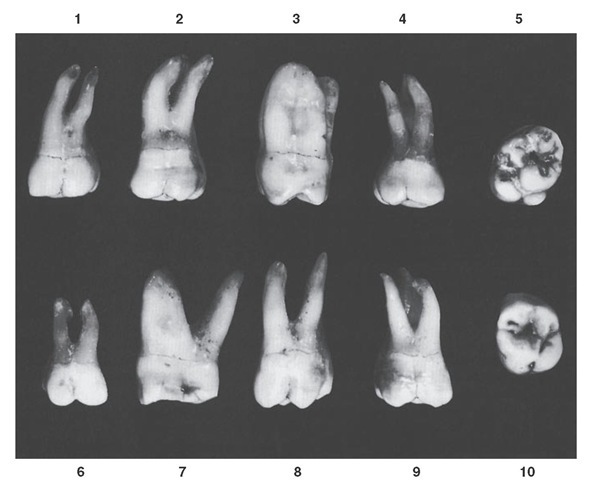
Figure 11-18 Maxillary first molar. Ten specimens with uncommon variations are shown. 1, Unusual curvature of buccal roots. 2, Roots abnormally long with extreme curvature. 3, Lingual and distobuccal roots fused. 4, Mesiodistal measurement of root trunk smaller than usual. 5, Extreme rhomboidal development of crown; fifth cusp with maximum development. 6, Tooth well developed but much smaller than usual. 7, Extreme buccolingual measurement. 8, Extreme length, especially of the distobuccal root; buccal cusps narrow mesiodistally. 9, Well-developed crown; roots poorly developed. 10, Extreme development of lingual portion of the crown compared with buccal development.
The four major cusps are well developed, with the small minor, or fifth, cusp appearing on the lingual surface of the mesiolingual cusp near the mesiolingual line angle of the crown. The fifth cusp may be indistinct, or all the cusp form may be absent. At this site, however, traces of developmental lines are nearly always present in the enamel.
The mesiolingual cusp is the largest cusp; it is followed in size by the mesiobuccal, distolingual, distobuccal, and fifth cusps.
If reduced to a geometric schematic figure, the occlusal aspect of this molar locates the various angles of the rhom-boidal figure as follows: acute angles, mesiobuccal and dis-tolingual; and obtuse angles, mesiolingual and distobuccal.
An analysis of the design of the occlusal surfaces of maxillary molars may be summarized here. Developmentally, only three major cusps can be considered as primary, the mesiolingual cusp and the two buccal cusps. The distolin-gual cusp is common to all of the maxillary molars; any other additional one, such as the cusp of Carabelli on first molars, must be regarded as secondary.
The triangular arrangement of cusps is reflected in the outline of the root trunks of maxillary molars when the teeth are sectioned in those areas.The distolingual cusp becomes progressively smaller on second and third maxillary molars, often disappearing as a major cusp (see Figure 11-11). Thus the triangular arrangement of the three important molar cusps is called the maxillary molar primary cusp triangle. The characteristic triangular figure, made by tracing the cusp outlines of these cusps, the mesial marginal ridge, and the oblique ridge of the occlusal surface, is representative of all maxillary molars.
The occlusal surface, or occlusal table as it is sometimes termed, of the maxillary first molar is within the confines of the cusp ridges and marginal ridges. The morphological features are now considered.
There are two major fossae and two minor fossae. The major fossae are the central fossa, which is roughly triangular and mesial to the oblique ridge, and the distal fossa, which is roughly linear and distal to the oblique ridge.
The two minor fossae are the mesial triangular fossa, immediately distal to the mesial marginal ridge, and the distal triangular fossa, immediately mesial to the distal marginal ridge (see Figure 11-l).
The oblique ridge is a ridge that crosses the occlusal surface obliquely. The union of the triangular ridge of the distobuccal cusp and the distal ridge of the mesiolingual cusp forms it. This ridge is reduced in height in the center of the occlusal surface, being about on a level with the marginal ridges of the occlusal surface. Sometimes it is crossed by a developmental groove that partially joins the two major fossae by means of its shallow sulcate groove.
The mesial marginal ridge and the distal marginal ridge are irregular ridges confluent with the mesial and distal cusp ridges of the mesial and distal major cusps.
The central fossa of the occlusal surface is a concave area bound by the distal slope of the mesiobuccal cusp, the mesial slope of the distobuccal cusp, the crest of the oblique ridge, and the crests of the two triangular ridges of the mesiobuccal and mesiolingual cusps. The central fossa has connecting sulci within its boundaries, with developmental grooves at the deepest portions of these sulci (sulcate grooves). In addition, it contains supplemental grooves, short grooves that are disconnected, and also the central developmental pit. A worn specimen may show developmental or sulcate grooves only.
In the center of the central fossa, the central developmental pit has sulcate developmental grooves radiating from it at obtuse angles to each other. This pit is located in the approximate center of that portion of the occlusal surface that is circumscribed by cusp ridges and marginal ridges (see Figure 11-1). From this pit the buccal developmental groove radiates buccally at the bottom of the buccal sulcus of the central fossa, continuing on to the buccal surface of the crown between the buccal cusps.
Starting again at the central pit, the central developmental groove is seen to progress in a mesial direction at an obtuse angle to the buccal sulcate groove. The central groove at the bottom of the sulcus of the central fossa usually terminates at the apex of the mesial triangular fossa. Here it is joined by short, supplemental grooves that radiate from its terminus into the triangular fossa. These supplemental grooves often appear as branches of the central groove. Occasionally one or more supplemental grooves cross the mesial marginal ridge of the crown.
The mesial triangular fossa is rather indistinct in outline, but it is generally triangular in shape, with its base at the mesial marginal ridge and its apex at the point where the supplemental grooves join the central groove.
An additional short developmental groove radiates from the central pit of the central fossa at an obtuse angulation to the buccal and central developmental grooves. Usually, it is considered a projection of one of these, because it is very short and usually fades out before reaching the crest of the oblique ridge. When it crosses the oblique ridge transversely, however, as it sometimes does, joining the central and distal fossae with a shallow groove, it is called the transverse groove of the oblique ridge (see Figure 11-17, 3, 4, and 5).
The distal fossa of the maxillary first molar is roughly linear in form and is located immediately distal to the oblique ridge. An irregular developmental groove traverses its deepest portion. This developmental groove is called the distal oblique groove. It connects with the lingual developmental groove at the junction of the cusp ridges of the mesiolin-gual and distolingual cusps. These two grooves travel in the same oblique direction to the terminus of the lingual groove, which is centered below the lingual root at the approximate center of the crown lingually (see Figure 11-5, LDG). If the fifth cusp development is distinct, a developmental groove outlining it joins the lingual groove near its terminus. Any part of the developmental groove that outlines a fifth cusp is called the fifth cusp groove.
The distal oblique groove in most cases shows several supplemental grooves. Two terminal branches usually appear, forming two sides of the triangular depression immediately mesial to the distal marginal ridge. These two sides, in combination with the slope mesial to the distal marginal ridge, form the distal triangular fossa. The distal outline of the distal marginal ridge of the crown shows a slight concavity.
The distolingual cusp is smooth and rounded from the occlusal aspect, and an outline of it, from the distal concavity of the distal marginal ridge to the lingual groove of the crown, describes an arch of an ellipse.
The lingual outline of the distolingual cusp is straight with the lingual outline of the fifth cusp, unless the fifth cusp is unusually large. In the latter case the lingual outline of the fifth cusp is more prominent lingually (see Figure 11-17, 9). The cusp ridge of the distolingual cusp usually extends lingually farther than the cusp ridge of the mesiolingual cusp.
Maxillary Second Molar
Figures 11-19 through 11-27 illustrate the maxillary second molar from all aspects. The maxillary second molar supple-ments the first molar in function. In describing this tooth, direct comparisons will be made with the first molar both in form and development.
Figure 11-19 Maxillary left second molar, buccal aspect. (Grid = 1 sq mm.)
Figure 11-20 Maxillary left second molar, lingual aspect. (Grid = 1 sq mm.)
Generally speaking, the roots of this tooth are as long as, if not somewhat longer than, those of the first molar (Table 11-2). The distobuccal cusp is not as large or as well developed, and the distolingual cusp is smaller. No fifth cusp is evident.
The crown of the maxillary second molar is 0.5 mm or so shorter cervico-occlusally than that of the first molar, but the measurement of the crown buccolingually is about the same.
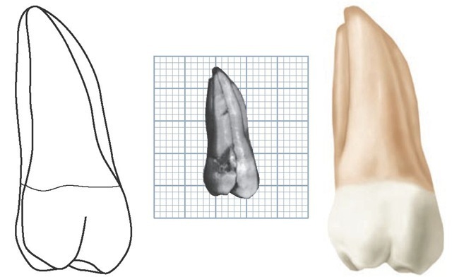
Figure 11-21 Maxillary left second molar, mesial aspect. (Grid = 1 sq mm.)
Figure 11-22 Maxillary left second molar, distal aspect. (Grid = 1 sq mm.)
Figure 11-23 Maxillary left second molar, occlusal aspect. (Grid = 1 sq mm.)
Figure 11-24 Maxillary second molar, buccal aspect. Ten typical specimens are shown.
Table 11-2 Maxillary Second Molar
Two types of maxillary second molars are found when the occlusal aspect is viewed: (1) The type that is seen most has an occlusal form that resembles that of the first molar, although the rhomboidal outline is more extreme. This is accentuated by the lesser measurement lingually. (2) The second type bears more resemblance to a typical third molar form. The distolingual cusp is poorly developed and makes the development of the other three cusps predominate. This results in a heart-shaped form from the occlusal aspect that is more typical of the maxillary third molar (see Figure 11-26, 1 and 7). Ten specimens with uncommon variations are shown in Figure 11-27.
