The mandibular premolars number four: two are situated in the right side of the mandible and two in the left side. They are immediately posterior to the mandibular canines and anterior to the molars.
The mandibular first premolars are developed from four lobes, as were the maxillary premolars. The mandibular second premolars are, in most instances, developed from five lobes, three buccal and two lingual lobes.
The first premolar has a large buccal cusp, which is long and well formed, with a small, nonfunctioning lingual cusp that in some specimens is no longer than the cingulum found on some maxillary canines (see Figure 10-10, 3 and 8; and Figure 10-12, 4 and 7). The second premolar has three well-formed cusps in most cases, one large buccal cusp and two smaller lingual cusps. The form of both mandibular premolars fails to conform to the implications of the term bicuspid, a term that implies two functioning cusps.
The mandibular first premolar has many of the characteristics of a small canine, because its sharp buccal cusp is the only part of it occluding with maxillary teeth. It functions along with the mandibular canine. The mandibular second premolar has more of the characteristics of a small molar, because its lingual cusps are well developed, a fact that places both marginal ridges high and produces a more efficient occlusion with antagonists in the opposite jaw. The mandibular second molar functions by being supplementary to the mandibular first molar.
The first premolar is always the smaller of the two mandibular premolars, whereas the opposite is true, in many cases, of the maxillary premolars.
Mandibular First Premolar
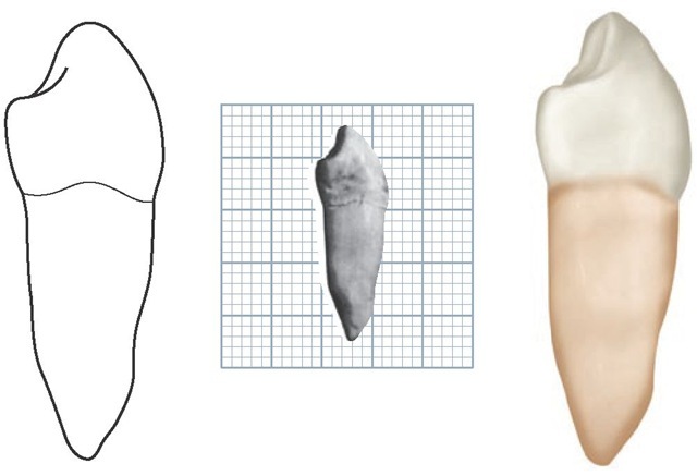
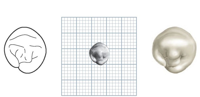
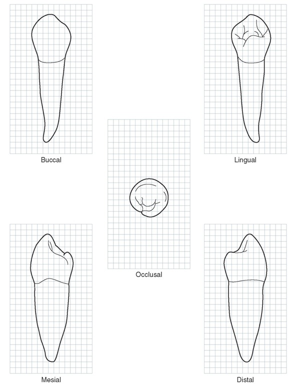
Figures 10-1 through 10-12 illustrate the mandibular first premolar from all aspects. The mandibular first premolar is the fourth tooth from the median line and the first posterior tooth in the mandible. This tooth is situated between the canine and second premolar and has some characteristics common to each of them.
The characteristics that resemble those of the mandibular canine are as follows:
1. The buccal cusp is long and sharp and is the only occluding cusp.
2. The buccolingual measurement is similar to that of the canine.
3. The occlusal surface slopes sharply lingually in a cervical direction.
4. The mesiobuccal cusp ridge is shorter than the distobuccal cusp ridge.
5. The outline form of the occlusal aspect resembles the outline form of the incisal aspect of the canine (compare Figure 10-6 and Figure 8-18).
The characteristics that resemble those of the second mandibular premolar are as follows:
1. Except for the longer cusp, the outline of crown and root from the buccal aspect resembles that of the second premolar.
2. The contact areas, mesially and distally, are near the same level.
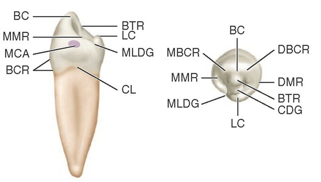
FIGURE 10-1 Mandibular right first premolar, mesial and occlusal aspects. BC, Buccal cusp; BTR, buccal triangular ridge; LC, lingual cusp; MLDG, mesiolingual developmental groove; CL, cervical line; BCR, buccal cervical ridge; MCA, mesial contact area; MMR, mesial marginal ridge; DBCR, distobuccal cusp ridge; DMR, distal marginal ridge; CDG, central developmental groove; MBCR, mesiobuccal cusp ridge.
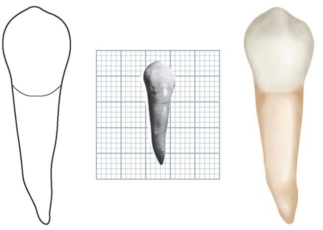
FIGURE 10-2 Mandibular right first premolar, buccal aspect. The specimen in this photograph shows a mesial inclination of the root. Mandibular premolars and canines have this tendency, although most of the roots of these teeth will curve, if at all, in a distal direction. (Grid = 1 sq mm.)
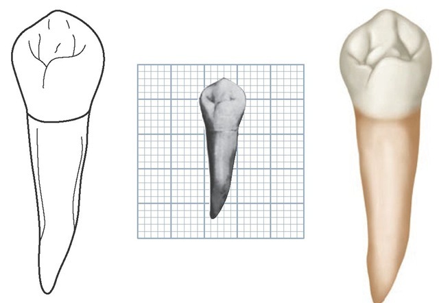
FIGURE 10-3 Mandibular right first premolar, lingual aspect. (Grid = 1 sq mm.)
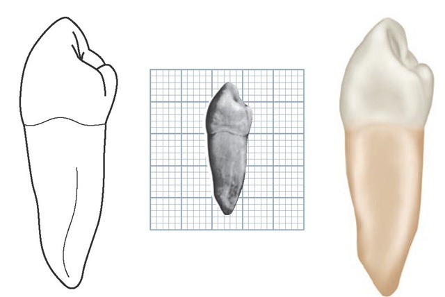
FIGURE 10-4 Mandibular right first premolar, mesial aspect. (Grid = 1 sq mm.)
FIGURE 10-5 Mandibular right first premolar, distal aspect. (Grid = 1 sq mm.)
FIGURE 10-6 Mandibular right first premolar, occlusal aspect. (Grid = 1 sq mm.)
Figure 10-7 Mandibular right first premolar. Graph outlines of five aspects are shown. (Grid = 1 sq mm.)
3. The curvatures of the cervical line mesially and distally are similar.
4. The tooth has more than one cusp.
Although the root of the mandibular first premolar is shorter generally than that of the mandibular second premolar, it is closer to the length of the second premolar root than it is to that of the mandibular canine (Table 10-1).
