Mandibular Third Molar
Figures 12-27 through 12-37 illustrate the mandibular third molar from all aspects.
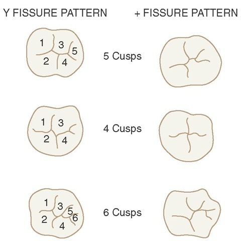
The mandibular third molar varies considerably in different individuals and presents many anomalies both in form and in position. It supplements the second molar in function, although the tooth is seldom as well developed, with the average mandibular third molar showing irregular development of the crown portion, with undersized roots, more or less malformed. However, its design usually conforms to the general plan of all mandibular molars, matching more closely the second mandibular molar in the number of cusps and occlusal design than it does the mandibular first molar.
Figure 12-27 Mandibular molar patterns in the right lower molar. Y and + fissure patterns are shown. 1, Protoconid; 2, metaconid; 3, hypoconid; 4, entoconid; 5, hypoconulid; 6, sixth cusp.
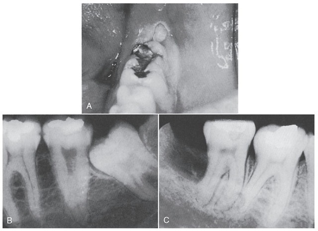
Figure 12-28 A, Partially erupted third molar. B, Impacted third molar. C, Defect immediately following extraction of a different third molar. Often the distal root surface of the second molar does not gain reattachment after a partially impacted third molar is extracted, especially if the surface has been exposed for some time prior to extraction.
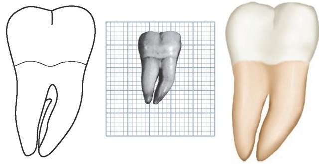
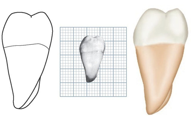
Figure 12-29 Mandibular right third molar, buccal aspect. (Grid = 1 sq mm.)
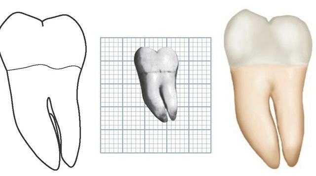
Figure 12-30 Mandibular right third molar, lingual aspect. (Grid = 1 sq mm.)
Figure 12-31 Mandibular right third molar, mesial aspect. (Grid = 1 sq mm.)
Figure 12-32 Mandibular right third molar, distal aspect. (Grid = 1 sq mm.)
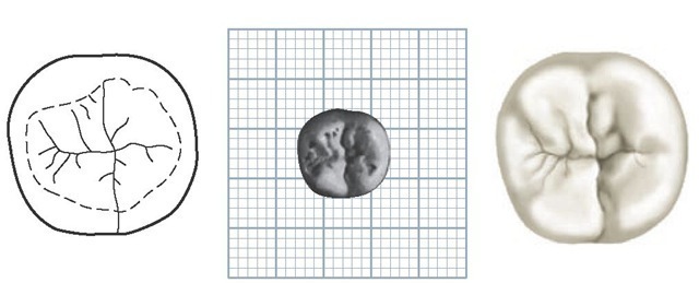
Figure 12-33 Mandibular right third molar, occlusal aspect. (Grid = 1 sq mm.)
Occasionally, mandibular third molars are seen that are well formed and comparable in size and development to the mandibular first molar.
Many instances of mandibular third molars with five or more cusps are found, with the crown portions larger than those of the second molar (Table 12-3). In these cases, the alignment and occlusion with other teeth is not normal, because insufficient room is available in the alveolar process of the mandible for the accommodation of such a large tooth, and the occlusal form is too variable.
Although it is possible to find dwarfed specimens of mandibular third molars (see Figure 12-37, 2), most of them that are not normal in size are larger than normal, in the crown portion particularly. Roots of these oversized third molars may be short and poorly formed.
The opposite situation is likely in maxillary third molars. Most of the anomalies are undersized. Mandibular third molars are the most likely to be impacted, wholly or partially, in the jaw. The lack of space accommodation is the chief cause.
If the third molar is congenitally absent from one side of the mandible or maxilla, it will most likely be absent from the other. However, no significant association is evident between third molar agenesis in the maxilla and mandible. Partial eruption of mandibular third molar teeth may result in periodontal defects on the distal aspects by the second molars and, in some instances, resorption of distal root surfaces occurs (see Figure 12-28). When third molars are to be restored, it should be remembered that the depth of the enamel on the occlusal surface is relatively greater than that on first or second molars.
DETAILED DESCRIPTION OF THE MANDIBULAR THIRD MOLAR FROM ALL ASPECTS
Buccal Aspect
From the buccal aspect, mandibular third molars vary considerably in outline. At the same time, they all have certain characteristics in common (see Figures 12-29 and 12-34).
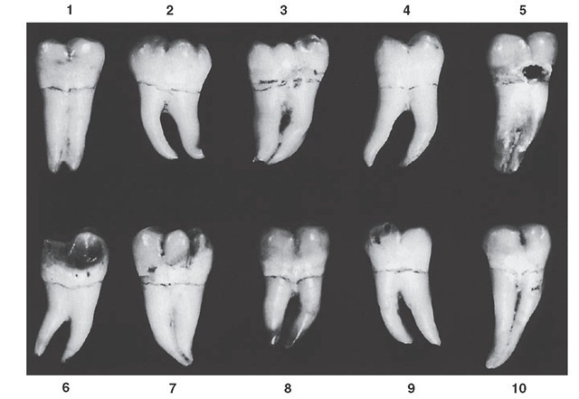
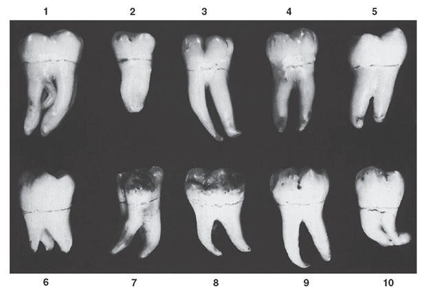
Figure 12-34 Mandibular third molar, buccal aspect. Ten typical specimens are shown.
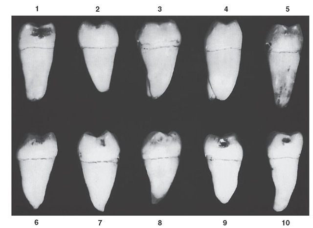
Figure 12-35 Mandibular third molar, mesial aspect. Ten typical specimens are shown.
The outline of the crowns from this aspect is in a general way that of all mandibular molars. The crown is wider at contact areas mesiodistally than at the cervix, the buccal cusps are short and rounded, and the crest of contour mesially and distally is located a little more than half the distance from cervical line to tips of cusps. The type of third molar, which is more likely to be in fair alignment and in good occlusion with other teeth, is the four-cusp type; this is smaller and shows two buccal cusps only from this aspect (see Figure 12-34, 1, 4, 5, 8, 9, and 10).
The average third molar also shows two roots, one mesial and one distal. These roots are usually shorter, with a poorer development generally, than the roots of first or second molars, and their distal inclination in relation to the occlusal plane of the crown is greater. The roots may be separated with a definite point of bifurcation, or they may be fused for all or part of their length (see Figure 12-34).
Figure 12-36 Mandibular third molar, occlusal aspect. Ten typical specimens are shown.
Figure 12-37 Mandibular third molar. Ten specimens with uncommon variations are shown. 1, Oversize generally, extra root lingually. 2, Dwarfed specimen; odd extra cusp; fused roots. 3, Crown resembling that of first molar; long, slender roots. 4, Formation closely resembling that of second molar. 5, Large crown; malformed roots. 6, Multicusp crown; dwarfed roots. 7, No resemblance to typical functional form. 8, Large crown; dwarfed roots. 9, Odd crown form and root form. 10, Crown long cervico-occlusally; roots fused and malformed.
Table 12-3 Mandibular Third Molar
|
First evidence of calcification |
|
8-10 yr |
|||||
|
Enamel completed |
12-16 yr |
||||||
|
Eruption |
17-21 yr |
||||||
|
Root completed |
18-25 yr |
||||||
|
Measurement Table |
|||||||
|
|
CERvico-occlusal Length of Crown |
Length of Root |
Mesìodìstal Mesìodìstal Dìameter of Dìameter Crown at of Crown Cervìx |
Labìo- or Buccolìngual Dìameter of Crown |
Labìo- or Buccolìngual Dìameter of Crown at CERvix |
Curvature of Cervìcal Lìne—Mesìal |
Curvature of Cervìcal Lìne—Dìstal |
|
Dimensions* suggested for carving technique |
7.0 |
11.0 |
10.0 7.5 |
9.5 |
9.0 |
1.0 |
0.0 |
Lingual Aspect
Observations from the lingual aspect add little to those already made from the buccal aspect. The mandibular third molar, when well developed, corresponds closely in form to the second molar except for size and root development (see Figure 12-30).
Mesial Aspect
From the mesial aspect, this tooth resembles the mandibular second molar except in dimensions (see Figures 12-31 and 12-35). The roots, of course, are shorter, with the mesial root tapering more from cervix to apex. The apex of the mesial root is usually more pointed.
Distal Aspect
The anatomical appearance of the distal portion of this tooth is much like that of the second molar except for size (see Figure 12-32).
Those specimens that have oversize crown portions are much more spheroidal above the cervical line. The distal root appears small, both in length and in buccolingual measurement, compared with the large crown portion.
Occlusal Aspect
The occlusal aspect of the third mandibular molar is quite similar to that of the second mandibular molar when the development is such as to facilitate good alignment and occlusion (see Figure 12-36, 2, 3, 4, and 6 through 9). The tendency is toward a more rounded outline and a smaller buccolingual measurement distally (see Figure 12-33).
