Detailed Description of the Mandibular Canine From All Aspects
Labial Aspect
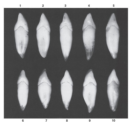
From the labial aspect, the mesiodistal dimensions of the mandibular canine are less than those of the maxillary canine. The difference is usually about 1 mm. The mandibular canine is broader mesiodistally than either of the mandibular incisors, for example, about 1 mm wider than the mandibular lateral incisor (see Figures 8-14, 8-19, 8-20, and 8-21).
The essential differences between mandibular and maxillary canines viewed from the labial aspect may be described as follows:
• The crowns of the mandibular canines appear longer. Sometimes they are longer, but the effect of greater length is emphasized by the narrowness of the crown mesiodistally and the height of the contact areas above the cervix.
• The mesial outline of the crown of the mandibular canine is nearly straight with the mesial outline of the root, with the mesial contact area being near the mesioincisal angle.
• When the cusp ridges have not been affected by wear, the cusp angle is on a line with the center of the root, as on the maxillary canine. The mesial cusp ridge is shorter.
• The distal contact area of the mandibular canine is more toward the incisal aspect than that of the maxillary canine but is not up to the level of the mesial area.
Figure 8-20 Mandibular right canine.
• The cervical line labially has a semicircular curvature apically.
• Many mandibular canines give the impression from this aspect of being bent distally on the root base. The maxillary canine crowns are more likely to be in line with the root.
• The mandibular canine root is shorter by l or 2 mm on average than that of the maxillary canine, and its apical end is more sharply pointed. Root curvatures are infrequent. When curvature of root ends is present, it is often in a mesial direction (see Figure 8-21, 1, 2, 3, and 4).
Lingual Aspect
In comparing the lingual aspect of the mandibular canine with that of the maxillary canine, the following differences are noted.
The lingual surface of the crown of the mandibular canine is flatter, simulating the lingual surfaces of mandibular incisors (see Figures 8-13, 8-15, 8-19, and 8-20). The cingulum is smooth and poorly developed. The marginal ridges are less distinct. This is true of the lingual ridge except toward the cusp tip, where it is raised. Generally speaking, the lingual surface of the crown is smooth and regular.
The lingual portion of the root is narrower relatively than that of the maxillary canine. It narrows down to little more than half the width of the labial portion.
Figure 8-21 Mandibular canine, labial aspect. Ten typical specimens are shown.
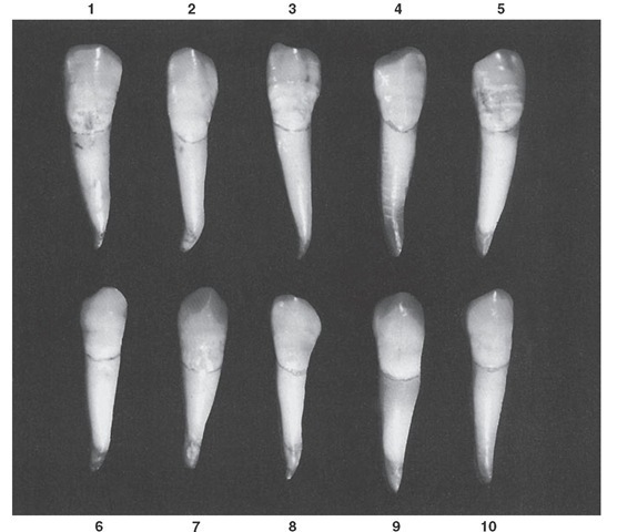
Figure 8-22 Mandibular canine, mesial aspect. Ten typical specimens are shown.
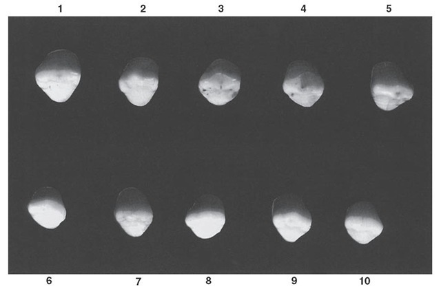
Figure 8-23 Mandibular canine, incisal aspect. Ten typical specimens are shown.
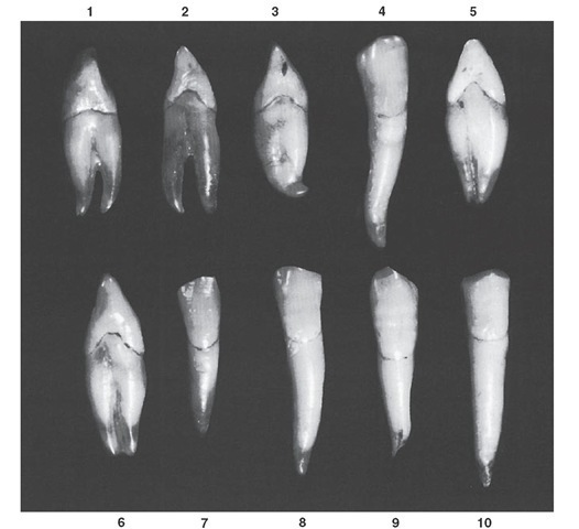
Figure 8-24 Mandibular canine. Ten specimens with uncommon variations are shown. 1, Well-formed crown; two roots, one lingual and one labial. 2, Same as specimen 1, with longer roots. 3, Well-formed crown portion; poorly formed root. 4, Root longer than average, with extreme curvature. 5, Deep developmental groove dividing the root. 6, Same as specimen 5. 7, Crown resembling mandibular lateral incisor; root short. 8, Root extra long, with odd mesial curvature starting at cervical third. 9, Crown extra long and irregular in outline. Root short and poorly formed at apex. 10, Crown with straight mesial and distal sides, wide at cervix, with a root of extreme length.
Table 8-2 Mandibular Canine
|
First evidence of calcification |
|
4-5 mo |
|||||
|
Enamel completed |
6-7 yr |
||||||
|
Eruption |
9-10 yr |
||||||
|
Root completed |
12-14 yr |
||||||
|
Measurement Table |
|||||||
|
Cervicoincisal Length of Crown |
Length of Root |
Mesiodistal Mesiodistal Diameter of Diameter Crown at of Crown Cervix |
Labio- or Buccolingual Diameter of Crown |
Labio- or Buccolingual Diameter of Crown at Cervix |
Curvature of Cervical Line—Mesial |
Curvature of Cervical Line—Distal |
|
|
Dimensions* suggested for carving technique |
11.0 |
16.0 |
7.0 5.5 |
7.5 |
7.0 |
2.5 |
1.0 |
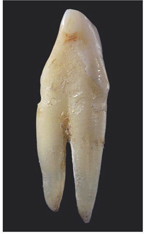
FIGURE 8-25 Mesial view of a left mandibular canine demonstrating prominent root bifurcation.
Mesial Aspect
From the mesial aspect, characteristic differences are evident between the two teeth in question. The mandibular canine has less curvature labially on the crown, with very little curvatu e directly above the cervical line. The curvature at the cervical portion as a rule is less than 0.5 mm. The lingual outline of the crown is curved in the same manner as that of the maxillary canine, but it differs in degree (see Figures 8-16, 8-19, 8-20, and 8-22).
The cingulum is not as pronounced, and the incisal portion of the crown is thinner labiolingually, which allows the cusp to appear more pointed and the cusp ridge to appear more slender. The tip of the cusp is more nearly centered over the root, with a lingual placement in some cases comparable to the placement of incisal ridges on mandibular incisors.
The cervical line curves more toward the incisal portion than does the cervical line on the maxillary canine.
The roots of the two teeth are quite similar from the mesial aspect with the possible exception of a more pointed root tip on the mandibular canine. The developmental depression mesially on the root of the mandibular canine is more pronounced and sometimes quite deep.
Distal Aspect
Little difference from the distal aspect can be seen between mandibular and maxillary canines except for those features mentioned under mesial aspect that are common to both (see Figures 8-17, 8-19, and 8-20).
Incisal Aspect
The outlines of the crowns of mandibular and maxillary canines from the incisal aspect are often similar (see Figures 8-13, 8-18, 8-19, 8-20, and 8-23). The main differences to be noted are as follows:
• The mesiodistal dimension of the mandibular canine is less than the labiolingual dimension. A similarity is evident in this, but the outlines of the mesial surface are less curved.
• The cusp tip and mesial cusp ridge are more likely to be inclined in a lingual direction in the mandibular canine, with the distal cusp ridge and the contact area extension distinctly so. Note that the cusp ridges of the maxillary canine with the contact area extensions are more nearly in a straight line mesiodistally from the incisal aspect.

