Detailed Description of the Maxillary Canine From All Aspects
Labial Aspect
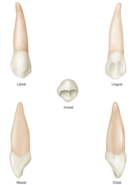
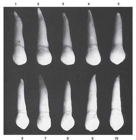
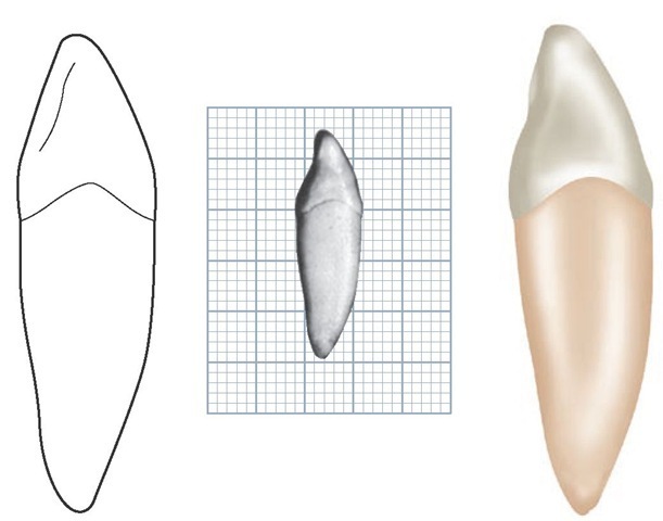
From the labial aspect, the crown and root are narrower mesiodistally than those of the maxillary central incisor. The difference is about 1 mm in most mouths. The cervical line labially is convex, with the convexity toward the root portion (see Figures 8-2, 8-7, 8-8, and 8-9).
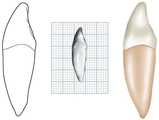
Figure 8-8 Maxillary right canine.
Mesially, the outline of the crown may be convex from the cervix to the center of the mesial contact area, or the crown may exhibit a slight concavity above the contact area from the labial aspect. The center of the contact area mesially is approximately at the junction of middle and incisal thirds of the crown.
Distally, the outline of the crown is usually concave between the cervical line and the distal contact area. The distal contact area is usually at the center of the middle third of the crown. The two levels of contact areas mesially and distally should be noted (see Figure 5-7, B and C).
Unless the crown has been worn unevenly, the cusp tip is on a line with the center of the root. The cusp has a mesial slope and a distal slope, the mesial slope being the shorter of the two. Both slopes show a tendency toward concavity before wear has taken place (see Figure 8-9, 5 and 6). These depressions are developmental in character.
The labial surface of the crown is smooth, with no developmental lines of note except shallow depressions mesially and distally, dividing the three labial lobes. The middle labial lobe shows much greater development than the other lobes. This produces a ridge on the labial surface of the crown. A line drawn over the crest of this ridge, from the cervical line to the tip of the cusp, is a curved one inclined mesially at its center. All areas mesial to the crest of this ridge exhibit convexity except for insignificant developmental lines in the enamel. Distally to the labial ridge (see incisal aspect), a tendency exists toward concavity at the cervical third of the crown, although convexity is noted elsewhere in all areas approaching the labial ridge (see Figure 8-11, 7, 8, and 9).
FIGURE 8-9 Maxillary canine, labial aspect. Ten typical specimens are shown.
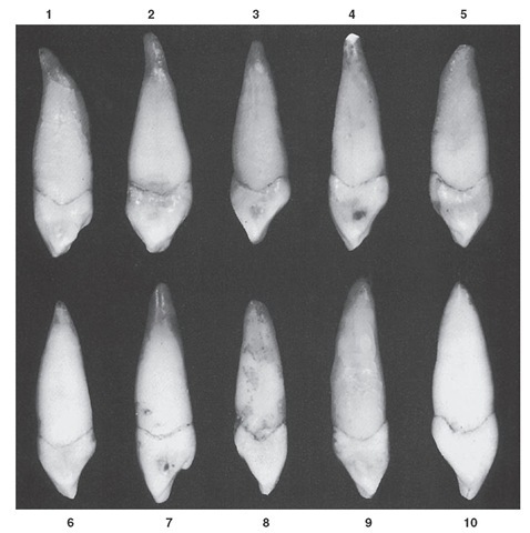
FIGURE 8-10 Maxillary canine, mesial aspect. Ten typical specimens are shown.
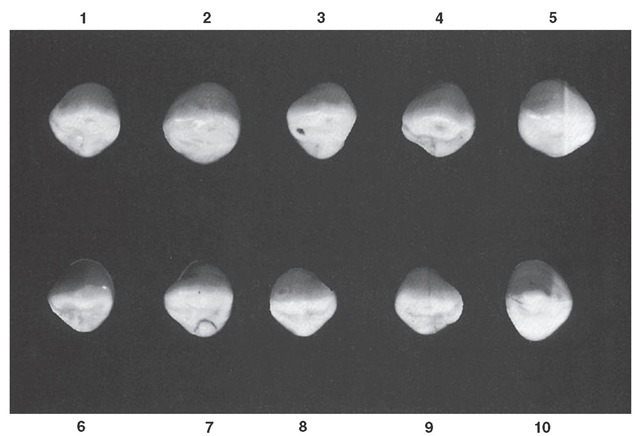
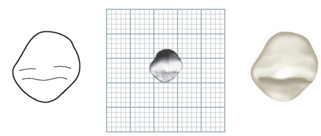
FIGURE 8-11 Maxillary canine, incisal aspect. Ten typical specimens are shown.
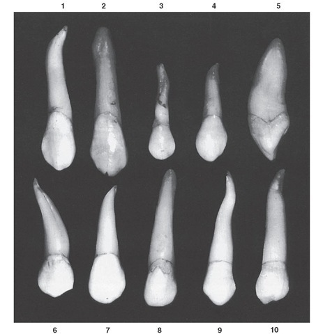
FIGURE 8-12 Maxillary canine. Ten specimens with uncommon variations are shown. 1, Crown very long, with extreme curvature at apical third of the root. 2, Entire tooth unusually long. Note hypercementosis at root end. 3, Very short crown; root small and malformed. 4, Mesiodistal dimension of crown at contact are extreme; calibration at cervix narrow in comparison; root short for crown of this size. 5, Extreme labiolingual calibration; root with unusual curvature. 6, Tooth malformed generally. 7, Large crown; short root. 8, Root overdeveloped and very blunt at apex. 9, Odd curvature of root; extra length. 10, Crown poorly formed; root extra long.
Table 8-1 Maxillary Canine
|
First evidence of calcification |
|
4-5 mo |
|||||
|
Enamel completed |
6-7 yr |
||||||
|
Eruption |
11-12 yr |
||||||
|
Root completed |
13-15 yr |
||||||
|
Measurement Table |
|||||||
|
|
Cervicoincisal Length of Crown |
Length of Root |
Mesiodistal Mesiodistal Diameter of Diameter Crown at of Crown Cervix |
Labio- or Buccolingual Diameter of Crown |
Labio- or Buccolingual Diameter of Crown at Cervix |
Curvature of Cervical Line—Mesial |
Curvature of Cervical Line—Distal |
|
Dimensions* suggested for carving technique |
10.0 |
17.0 |
7.5 5.5 |
8.0 |
7.0 |
2.5 |
1.5 |
The root of the maxillary canine appears slender from the labial aspect when compared with the bulk of the crown; it is conical in form with a bluntly pointed apex. It is not uncommon for this root to have a sharp curve in the vicinity of the apical third. This curvature may be in a mesial or distal direction, but in most instances is the latter (see Figure 8-9, 1 and 6). The labial surface of the root is smooth and convex at all points.
Lingual Aspect
The crown and root are narrower lingually than labially.
The cervical line from the lingual aspect differs somewhat from the curvature found labially and shows a more even curvature. The line may be straight for a short interval at this point (see Figures 8-3, 8-7, and 8-8).
The cingulum is large and, in some instances, is pointed like a small cusp (see Figure 8-10, 7). In this type, definite ridges are found on the lingual surface of the crown below the cingulum and between strongly developed marginal ridges. Although depressions are to be found between these ridge forms, seldom are any deep developmental grooves present.
Occasionally, a well-developed lingual ridge is seen that is confluent with the cusp tip; this extends to a point near the cingulum. Shallow concavities are evident between this ridge and the marginal ridges. When these concavities are present, they are called mesial and distal lingual fossae (see Figures 8-1 and 8-8).
Sometimes the lingual surface of the canine crown is so smooth that fossae or minor ridges are difficult to distinguish. However, a tendency exists toward concavities where the fossae are usually found, and heavy marginal ridges with a well-formed cingulum are to be expected. The smooth cingulum, marginal ridges, and lingual portion of the incisal ridges are usually confluent, with little evidence of developmental grooves.
The lingual portion of the root of the maxillary canine is narrower than the labial portion. Because of this formation,much of the mesial and distal surface of the root is visible from the lingual aspect. Developmental depressions mesially and distally may be seen on most of these roots, extending most of the root length. The lingual ridge of the root is rather narrow, but it is smooth and convex at all points from the cervical line to the apical end (see Figure 8-3).
Mesial Aspect
The mesial aspect of the maxillary canine presents the outline of the functional form of an anterior tooth. However, it generally shows greater bulk and greater labiolingual measurement than any of the other anterior teeth (see Figures 8-4, 8-7, 8-8, and 8-10).
The outline of the crown is wedge-shaped, the greatest measurement being at the cervical third and the wedge point being represented by the tip of the cusp.
The curvature of the crown below the cervical line labi-ally and lingually corresponds in extent to the curvature of the maxillary central and lateral incisors. Nevertheless, the crest of that curvature is found at a level more incisal, because the middle labial and the lingual lobes are more highly developed (see Figure 8-10, 5 and 10). Many canines show a flattened area labially at the cervical third of the crown, which appears as a straight outline from the mesial aspect. It is questionable just how much wear has to do with this effect (see Figure 8-10, 1 and 2).
Below the cervical third of the crown, the labial face may be presented by a line only slightly convex from the crest of curvature at the cervical third to the tip of the cusp. The line usually becomes straighter as it approaches the cusp.
The entire labial outline from the mesial aspect exhibits more convexity from the cervical line to the cusp tip than the maxillary central incisor does from cervix to incisal edge.
The lingual outline of the crown from the mesial aspect may be represented by a convex line describing the cingu-lum, which convexity straightens out as the middle third is reached, becoming convex again in the incisal third (see Figure 8-10, 10).
The cervical line that outlines the base of the crown from this aspect curves toward the cusp on average by approximately 2.5 mm at the cementoenamel junction. The outline of the root from this aspect is conical, with a tapered or bluntly pointed apex. The root may curve labially toward the apical third. The labial outline of the root may be almost perpendicular, with most of the taper appearing on the lingual side (see Figure 8-10, 4 and 9).
The position of the tip of the cusp in relation to the long axis of the root is different from that of the maxillary central and lateral incisors. Although the specimen illustrations in Figures 8-4 and 8-5 do not show this difference, most specimens in Figure 8-10 show it conclusively. A line bisecting the cusp is labial to a line bisecting the root. Lines bisecting the roots of central and lateral incisors also bisect the incisal ridges.
The mesial surface of the canine crown presents convexities at all points except for a small, circumscribed area above the contact area, where the surface is concave and flat between that area and the cervical line.
The mesial surface of the root appears broad, with a shallow developmental depression for part of the root length. Developmental depressions on the heavy roots help anchor the teeth in the alveoli and help prevent rotation and displacement.
Distal Aspect
The distal aspect of the maxillary canine shows somewhat the same form as the mesial aspect, with the following variations: the cervical line exhibits less curvature toward the cusp ridge; the distal marginal ridge is heavier and more irregular in outline; the surface displays more concavity, usually above the contact area; and the developmental depression on the distal side of the root is more pronounced (see Figures 8-5, 8-7, and 8-8).
Incisal Aspect
The incisal aspect of the maxillary canine emphasizes the proportions of this tooth mesiodistally and labiolingually (see Figures 8-6, 8-7, 8-8, and 8-11). In general, the labiolingual dimension is greater than the mesiodistal. Occasionally, the two measurements are about equal (see Figure 8-11, 8). Other instances appear in which the crown is larger than usual in a labiolingual direction (see Figure 8-11, 10).
From the incisal aspect, if the tooth is correctly posed so that the long axis of the root is directly in the line of vision, the tip of the cusp is labial to the center of the crown labio-lingually and mesial to the center mesiodistally.
If the tooth were to be sectioned labiolingually beginning at the center of the cusp of the crown, the two sections would show the root rather evenly bisected, with the mesial portion carrying a narrower portion of the crown mesiodis-tally than that carried by the distal section of the tooth. (Note the proportions demonstrated by the fracture line in the enamel in Figure 8-11, 9.) Nevertheless, the mesial section shows a crown portion with greater labiolingual bulk. The crown of this tooth gives the impression of having the entire distal portion stretched to make contact with the first premolar.
The ridge of the middle labial lobe is very noticeable labially from the incisal aspect. It attains its greatest convexity at the cervical third of the crown, becoming broader and flatter at the middle and incisal thirds.
The cingulum development makes up the cervical third of the crown lingually. A shorter arc than the one labially from this aspect may describe the outline of the cingulum. This comparison coincides with the relative mesiodistal dimensions of the root lingually and labially.
A line bisecting the cusp and cusp ridges drawn in the mesiodistal direction is almost always straight and bisects the short arcs representative of the mesial and distal contact areas. This fact emphasizes the close relation between maxillary canines and some lateral incisors, since they resemble each other in this characteristic (compare Figure 8-11, 7 with Figure 6-18, 1).The latter are supposed to be in the majority. Naturally, the lateral incisors that resemble canines are relatively wide labiolingually, and those resembling central incisors are narrow in that direction.
The incisal aspect of most canines, maxillary or mandibular, may be outlined in many cases by a series of arcs. Figure 8-11, 6, for example, could be drawn almost perfectly with the aid of a French curve, a drawing instrument used in drafting to draw arcs of varying degrees.
Mandibular Canine
Figures 8-13 through 8-24 illustrate the mandibular canine in various aspects. Because maxillary and mandibular canines bear a close resemblance to each other, direct comparisons are made with the maxillary canine in describing the mandibular canine.
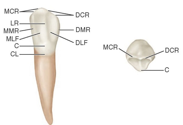
FIGURE 8-13 Mandibular right canine, lingual and incisal aspects. MCR, Mesial cusp ridge; LR, lingual ridge; MMR, mesial marginal ridge; MLF, mesiolingual fossa; C, cingulum; CL, cervical line; DLF, distolingual fossa; DMR, distal marginal ridge; DCR, distal cusp ridge.
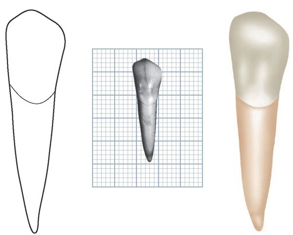
FIGURE 8-14 Mandibular left canine, labial aspect. (Grid = 1 sq mm.)
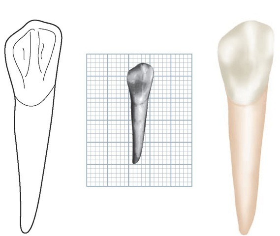
FIGURE 8-15 Mandibular left canine, lingual aspect. (Grid = 1 sq mm.)
FIGURE 8-16 Mandibular left canine, mesial aspect. (Grid = 1 sq mm.)
FIGURE 8-17 Mandibular left canine, distal aspect. (Grid = 1 sq mm.)
FIGURE 8-18 Mandibular left canine, incisal aspect. (Grid = 1 sq mm.)
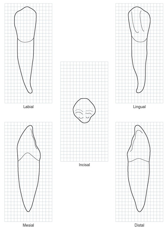
The mandibular canine crown is narrower mesiodistally than that of the maxillary canine, although it is just as long in most instances and in many instances is longer by 0.5 to 1 mm (Table 8-2). The root may be as long as that of the maxillary canine, but usually it is somewhat shorter. The labiolingual diameter of crown and root is usually a fraction of a millimeter less.
The lingual surface of the crown is smoother, with less cingulum development and less bulk to the marginal ridges. The lingual portion of this crown resembles the form of the lingual surfaces of the mandibular lateral incisors.
The cusp of the mandibular canine is not as well developed as that of the maxillary canine, and the cusp ridges are thinner labiolingually. Usually the cusp tip is on a line with the center of the root, from the mesial or distal aspect, but sometimes it lies lingual to the line, as with the mandibular incisors.
A variation in the form of the mandibular canine is bifurcated roots. This variation is not rare (see Figure 8-24, 1, 2, 5, 6 and Figure 8-25).
Figure 8-19 Mandibular right canine. Graph outlines of five aspects are shown. (Grid = 1 sq mm.)
