Crown and Root Fractures
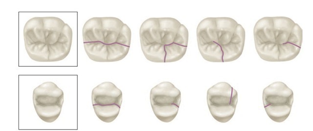
Fractures of teeth may involve the crown, the crown and root, or the root. Probably the most common are fractures of the crown.3 In some cracked teeth, only the enamel is involved; in others, both the enamel and dentin may be affected; but initially or later, the pulp also may be involved with or without the loss of tooth structure (e.g., cusp). In severe trauma the whole crown may be lost. The clinician should be familiar with the most likely places, morphologically, for cracking or fractures to occur. The most likely places involve developmental grooves (Figure 13-26), often in relation to restorations.
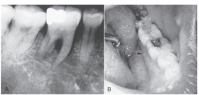
Fractures of cusps of the maxillary first premolar and mandibular first molars often occur along developmental grooves or stress lines as shown in Figure 13-26. Such fractures may occur in connection with bruxism and clenching. It is not uncommon for the distolingual cusp of the mandibular first molar to fracture in association with a large restoration, which leads to pulpitis (Figure 13-27).
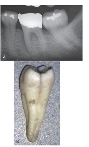
Fractures of the root cause symptoms that resemble those of other dental problems; therefore the diagnosis of root fracture may be difficult,3 especially when the fracture does not appear on the radiograph (Figure 13-28).
Fractures may be an extension from the crown and may extend to the pulp or involve only the root adjacent to the periodontal ligament. Horizontal root fractures are most likely the result of external physical trauma or clenching and bruxism. Vertical root fractures may be caused by bruxing and clenching or may be the result of restorations with endodontic posts in root canals.
RELATION OF POSTERIOR ROOT APICES TO THE MANDIBULAR CANAL
Of interest to the clinician doing endodontics, periapical surgery, or placement of implants is the position of the mandibular nerve (canal) and the mental foramen.
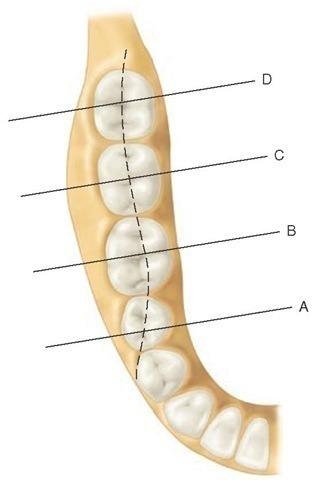
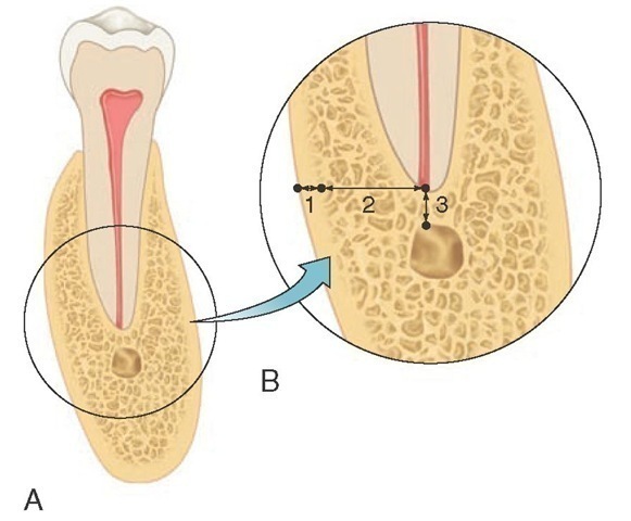
The mandibular nerve traversing the mandible in the mandibular canal occupies different positions relative to the apices of the molar and premolar teeth (Figure 13-29, A through D). In Figure 13-30, the distance from the outer to the inner surface of the buccal plate (1), from the inner surface of the cortical plate to the apex of a tooth (2), and from the canal to the apex of the tooth (3) reflect the data obtained from sections of the mandible.4
The mean width of the buccal plate (Figure 13-30, B, 1) over the premolar is 1.9 ± 0.49 mm; over the first molar, 2.38 ± 0.57 mm; over the second molar, 5.6 ± 0.93 mm; and over the third molar, 2.34 ± 1.0 mm.
The mean distance from the inner buccal plate to the apex of the tooth (Figure 13-30, B, 2) for the second premolar is 3.78 ± 1.04 mm; for the first molar, 4.1 ± 0.98 mm; for the second molar, 7.1 ± 1.4 mm; and for the third molar, 4.03 ± 1.8 mm.
The mean vertical distance from the canal to the apex (Figure 13-30, B, 3) for the second premolar is 3.07 ± 0.43 mm; for the first molar, 4.03 ± 0.31 mm; for the second molar, 2.5 ± 0.25 mm; and for the third molar, 1.96 ± 0.27 mm.
With regard to the buccolingual alignment of the mandibular canal to root apices, the canal is in vertical alignment with the second premolars for 65% of the time, slightly lingual to the apices of the first molar apices for 71% of the time, in line with the apices of second molars for 73% of the time, and in line with the third molars for 56% of the time.
Data are based on the vertical axis of each tooth with the recognition that more than one apex is present. Thus, the position of the mandibular canal relative to the apices of the teeth tends to follow the dotted line in Figure 13-29.
The position of the mental foramen relative to the mandibular premolars and first molar can be described as follows: the foramen is between the first and second premolars 33% of the time, in line with the second premolar 11% of the time, and distal to the second premolar 56% of the time. In terms of vertical position, the foramen can be found coronal to the apices 22% of the time, at the apices 15% of the time, and below the apices 63% of the time.4
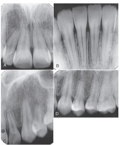
Figure 13-25 A, Large pulp chambers and pulp canals in a young adult. B, Mandibular incisors. Normal pulp canals. C, Lateral incisor and canine. Normal chambers and canals. Root resorption on lateral incisor. D, Maxillary canine and premolars with normal chambers and canals. Root resorption on first premolar associated with orthodontics.
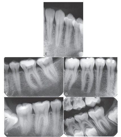
Figure 13-25 cont’d E, Mandibular lateral incisor, canine, and first premolar with normal chambers and canals. F, Mandibular second premolar and first and second molars with normal chambers and canals. Note curvature of the mesial root of the first molar. G, Mandibular molars and premolars with normal chambers and pulp canals. H, Impacted third molar adjacent to second mandibular molar. The pulp chambers and pulp canals are normal. I, First and second primary molars being resorbed in association with the erupting permanent premolars. Mandibular second molar in the process of erupting.
Figure 13-26 Fracture lines most commonly seen in first maxillary premolar and first mandibular molar.
Figure 13-27 A, Radiograph showing periapical radiolucency associated with parulis shown in B. B, Fracture of distolingual cusp related to undermined tooth structure and amalgam restoration.
Figure 13-28 A, Radiograph showing loss of lamina dura on the mesial root of the mandibular molar but no indication of root fracture. B, Longitudinal fracture on the distal aspect of the crown and coronal root.
Figure 13-29 Diagrammatic representation of sections through the mandible at the position of the second premolar and first, second, and third molars. Dotted line represents the faciolingual position of the mandibular canal.
Figure 13-30 A, Diagrammatic representation of a cross section of the mandible. B, Enlarged view with three measurements taken as indicated in the text.
