Facial and Lingual Relations of Each Tooth in One Arch to Its Antagonists in the Opposing Arch in Centric Occlusion
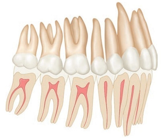
In the intercuspal position, facial views of the normal dentition show each tooth of one arch in occlusal with portions of two others in the opposing arch (Figure 16-24; see also Figure 16-20) with the exception of the mandibular central incisors and the maxillary third molars. Each of the exceptions named has one antagonist only in the opposing jaw.
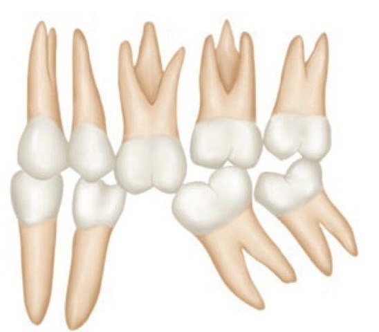
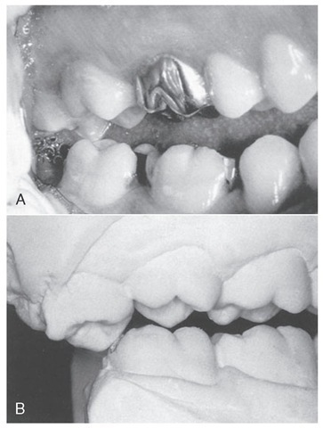
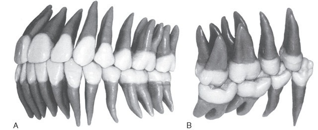
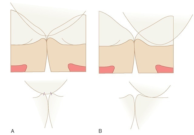
Because each tooth has two antagonists, the loss of one still leaves one antagonist remaining, which will keep the tooth in occlusal contact with the opposing arch and keep it in its own arch relation at the same time by preventing elongation and displacement through the lack of antagonism. The permanency of the arch forms depends on the mutual support of the teeth in contact with each other. When a tooth is lost, the adjoining teeth in the same arch may, depending on occlusal forces, migrate in an effort to fill the void (Figure 16-25). The migration of adjoining teeth disturbs the contact relationships in that vicinity. In the meantime, tooth movement changes occlusal relations with antagonists in opposing dental arches. The common result is hypereruption of the tooth opposing the space left by the lost tooth (Figure 16-26).
Figure 16-24 Schematic drawing showing the facial (buccal) relations of teeth in opposing arches in an idealized occlusion.
Figure 16-25 Illustration demonstrating possible migration, extrusion, and improper contacts and occlusal relations following loss of a mandibular molar.
Occlusal Contacts and Intercuspal Relations Between Arches
A working knowledge of occlusal contact and intercusp relations of both dental arches in the intercuspal position or centric occlusion is necessary for any discussion of occlusal relations, whether for the natural dentition or a proposed restoration of the dentition. Thus the dentist should know for discussion purposes where a particular supporting cusp makes contact with a centric stop on the opposing tooth. For example, the lingual cusps of the maxillary posterior teeth and the buccal cusps of the posterior mandibular teeth are referred to as supporting cusps.
Areas of occlusal contact that a supporting cusp makes with opposing teeth in centric occlusion are centric stops. The area of contact on the supporting cusp that makes contact with the opposing tooth in centric occlusion is also a centric stop. Therefore centric stops are areas of a tooth that make contact with opposing teeth in the intercuspal position (centric occlusion) and contribute to occlusal stability.
Figure 16-26 A, Hypererupted third molar due to loss of mandibular third molar. B, Casts mounted in centric relation on articulator showing undesirable centric relation contact of extruded molar with the lower molar.
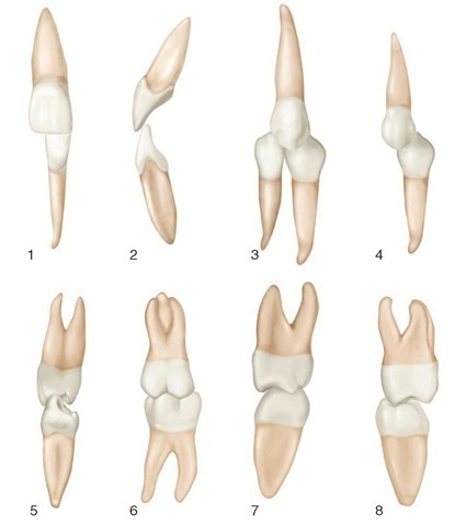
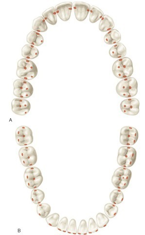
Thus, for example, the mesiolingual cusp of the maxillary first molar (a supporting cusp) makes contact with the central fossa (central stop) of the mandibular first molar (Figures 16-27 and 16-28, first molar views6-8). The clinical application of the occlusal scheme of contacts shown in Figure 16-27 is based on the concept of obtaining occlusal stability (e.g., in the intercuspal position, as in clenching, occlusal forces should be directed along the long axes of the teeth). An idealized schematic representation of all centric stops is shown in Figure 16-29.
Lingual views of the occlusal relations with the teeth in centric occlusion (Figure 16-30) show the intercuspation of lingual cusps and how far the maxillary teeth occlude laterally to the lingual cusps of the mandibular arch (Figure 16-31).
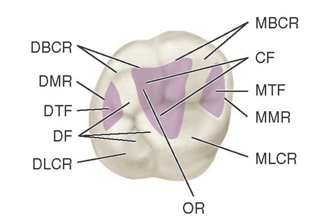
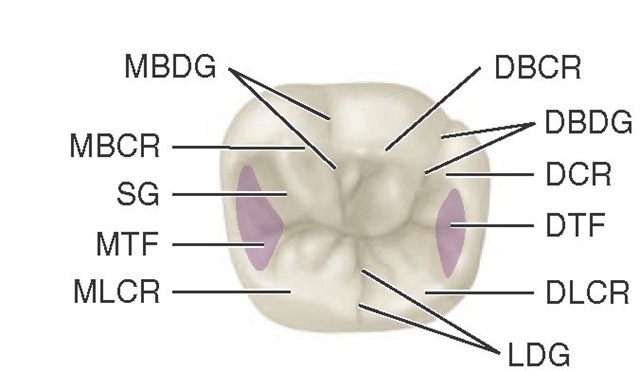
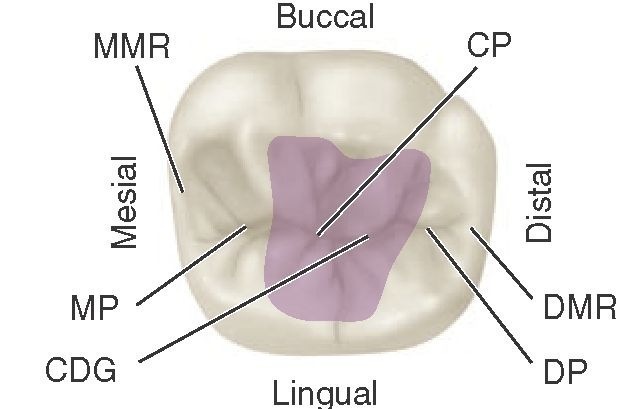
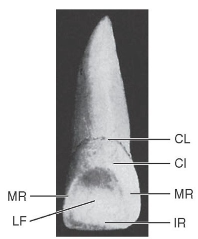
Figures and legends featuring landmarks on teeth are repeated here for use as a ready reference for identifying occlusal contacts (Figures 16-32 through 16-35).
Cusp, Fossa, and Marginal Ridge Relations
The contact relationship of the supporting cusps of the molars and premolars and fossae of the opposing teeth is shown in Figure 16-36.
Figure 16-27 Example of idealized cusp-fossa relationship. A, Mesiolingual cusp of maxillary first molar occludes in the central fossa of the mandibular first molar. Distal buccal cusp of mandibular first molar occludes in the central fossa of the maxillary first molar. B, Concept of occlusion in which all supporting cusps occlude in fossae.
Figure 16-28 Normal intercuspation of maxillary and mandibular teeth. 1, Central incisors (labial aspect). 2, Central incisors (mesial aspect). 3, Maxillary canine in contact with mandibular canine and first premolar (facial aspect). 4, Maxillary first premolar and mandibular first premolar (buccal aspect). 5, Maxillary first premolar and mandibular first premolar (mesial aspect). 6, First molars (buccal aspect). 7, First molars (mesial aspect). 8, First molars (distal aspect).
Figure 16-29 Idealized scheme for all contacts of supporting cusps with fossae and marginal ridges of opposing teeth. Such contact relations on all teeth are seldom found in the natural dentition. A, Maxillary arch. B, Mandibular arch.
The simulated relationship does not reflect all the variance that may occur in these relationships. The lingual cusps of the maxillary premolars do not necessarily make contact in the fossa of the mandibular but occlude with the marginal ridges of the premolars or premolars and first molars as indicated in Figures 16-36, A, and 16-37.
Figure 16-30 Lingual view of the teeth in centric occlusion.
Concept of 138 Points of Occlusal Contact
One scheme of occlusal contacts presented by Hellman included 138 points of possible occlusal contacts for 32 teeth.8 Later, with some modifications for application to complete occlusal restoration, most of the same contacts were made a part of the concept of occlusion in which supporting cusps and opposing stops (in centric relation) are tripoded and, with lateral/protrusive movements, immediate disocclusion of the posterior teeth takes place with canine (cuspid) guidance.
The list of occlusal contacts (total, 138) follows:
1. Lingual surfaces of maxillary incisors and canines, 6
2. Labial surface of mandibular incisors and canines, 6
3. Triangular ridges of maxillary buccal cusps of premolars and molars, 16
4. Triangular ridges of lingual cusps of mandibular premolars and molars, 16
5. Buccal embrasure of mandibular premolars and molars, 8
Figure 16-31 Occlusal contact relations in the intercuspal position consistent with Angle Class I molar relationship. A, Viewed from the facial aspect. B, Viewed from the lingual aspect.
Figure 16-32 Maxillary right first molar, occlusal landmarks. MBCR, Mesiobuccal cusp ridge; CF, central fossa (shaded area); MTF, mesial triangular fossa (shaded area); MMR, mesial marginal ridge; MLCR, mesiolingual cusp ridge; OR, oblique ridge; DLCR, distolingual cusp ridge; DF, distal fossa; DTF, distal triangular fossa (shaded area); DMR, distal marginal ridge; DBCR, distobuccal cusp ridge.
Figure 16-33 Mandibular right first molar, occlusal aspect. DBCR, Distobuccal cusp ridge; DBDG, distobuccal developmental groove; DCR, distal cusp ridge; DTF, distal triangular fossa (shaded area); DLCR, distolingual cusp ridge; LDG, lingual developmental groove; MLCR, mesiolingual cusp ridge; MTF, mesial triangular fossa (shaded area); SG, supplemental groove; MBCR, mesiobuccal cusp ridge; MBDG, mesiobuccal developmental groove.
Figure 16-34 Mandibular right first molar, occlusal aspect. Shaded area is the central fossa. CP, Central pit; DMR, distal marginal ridge; DP, distal pit; CDG, central developmental groove; MP, mesial pit; MMR, mesial marginal ridge.
Figure 16-35 Maxillary central incisor (lingual aspect). CL, Cervical line; CI, cingulum; MR, marginal ridge; IR, incisal ridge; LF, lingual fossa.
Figure 16-36 A, Relationship of supporting cusps to marginal ridges. B, relationship of supporting cusps to fossae.
6. Lingual embrasures of maxillary premolars and molars (including the canine and first premolar embrasure accommodating the mandibular premolar), 10
7. Lingual cusp points of maxillary premolars and molars, 16
8. Buccal cusp points of mandibular premolars and molars, 16
9. Distal fossae of premolars, 8
10. Central fossae of the molars, 12
11. Mesial fossae of the mandibular molars, 6
12. Distal fossae of the maxillary molars, 6
13. Lingual grooves of the maxillary molars, 6
14. Buccal grooves of the mandibular molars, 6*
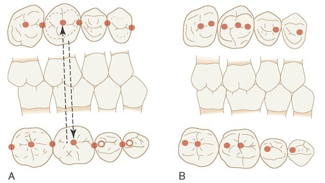
Therefore, if a complete description without the omission of any detail of ideal occlusion is desired, close scrutiny of a good skull or casts showing 32 teeth would make it possible to compile a list of all ridge-sulcus combinations, all cusp embrasure combinations, and so on. Usually, if the combinations of points that have been mentioned in the last few pages can be established, some details such as the approximate location of hard contact in occlusion are automatic. Friel’s concept of occlusal contacts in an "ideal" occlusion is illustrated in Figure 16-37.43 Compare with the contacts in Figure 16-29.
Concepts of ideal occlusion are used primarily in orthodontics and restorative dentistry. Application of the goal of a total of 138 points for orthodontics and complete oral restorative rehabilitation has not been shown to be practical or necessary for occlusal stability or function. That is not to say that such concepts for ideal occlusal contacts have no value.
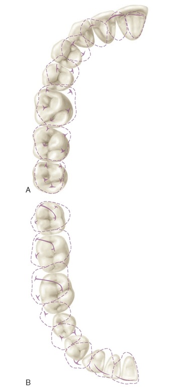
Figure 16-37 Contact relations in the intercuspal position (centric occlusion). A, Maxillary teeth with dotted lines superimposed on mandibular teeth. Heavy lines and T shapes within dotted lines denote ridges and cusp tips. B, Mandibular teeth, with dotted lines of maxillary teeth superimposed in occlusion. Note the slanted heavy lines of maxillary molars that mark the shape and location of oblique ridges.
Reasonable application of an idealized cusp-fossa relationship as indicated in Figures 16-27 and 16-37 are applicable to clinical dentistry. A complete set of the contacts in Figure 16-37 is not a common feature of the natural dentition.
Figure 16-38 Projected protrusive (P), working (W), and balancing (B) side paths on maxillary and mandibular first molars made by supporting cusps, that is, mesiolingual cusp of the maxillary molar projected on the mandibular molar and distobuccal cusp of the mandibular molar on the maxillary molar. D, Distal; M, mesial.
