Macromolecules of the cell
The six basic molecules are used by all cells to construct five essential macromolecules: proteins, RNA, DNA, phospholipids, and polysaccharides. Macromolecules have primary, secondary, and tertiary structural levels. The primary structural level refers to the chain that is formed by linking the building blocks together. The secondary structure involves the bending of the linear chain to form a three-dimensional object. Tertiary structural elements involve the formation of chemical bonds between some of the building blocks in the chain to stabilize the secondary structure. A quaternary structure can also occur when two identical molecules interact to form a dimer or double molecule.
Proteins are long chains or polymers of amino acids. The primary structure is held together by peptide bonds that link the car-boxyl end of one amino acid to the amino end of a second amino acid. Thus once constructed, every protein has an amino end and a carboxyl end. An average protein consists of about 400 amino acids. There are 21 naturally occurring amino acids; with this number the cell can produce an almost infinite variety of proteins. Evolution and natural selection, however, have weeded out most of these, so that eukaryote cells function well with 10,000 to 30,000 different proteins.
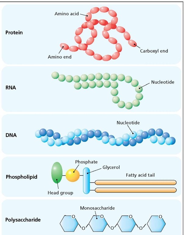
Macromolecules of the cell. Protein is made from amino acids linked together to form a long chain that can fold up into a three-dimensional structure. RNA and DNA are long chains of nucleotides. RNA is generally single-stranded, but can form localized double-stranded regions. DNA is a double-stranded helix, with one strand coiling around the other. A phospholipid is composed of a hydrophilic head-group, a phosphate, a glycerol molecule and two hydrophobic fatty acid tails. Polysaccharides are sugar polymers.
In addition, this select group of proteins has been conserved over the past 2 billion years (i.e., most of the proteins found in yeast can also be found, in modified form, in humans and other higher organisms) The secondary structure of a protein depends on the amino acid sequence and can be quite complicated, often producing three-dimensional structures possessing multiple functions.
RNA is a polymer of the ribonucleotides adenine, uracil, cytosine and guanine. RNA is generally single stranded, but it can form localized double-stranded regions by a process known as complementary base pairing, whereby adenine forms a bond with uracil and cytosine pairs with guanine. RNA is involved in the synthesis of proteins and is a structural and enzymatic component of ribosomes.
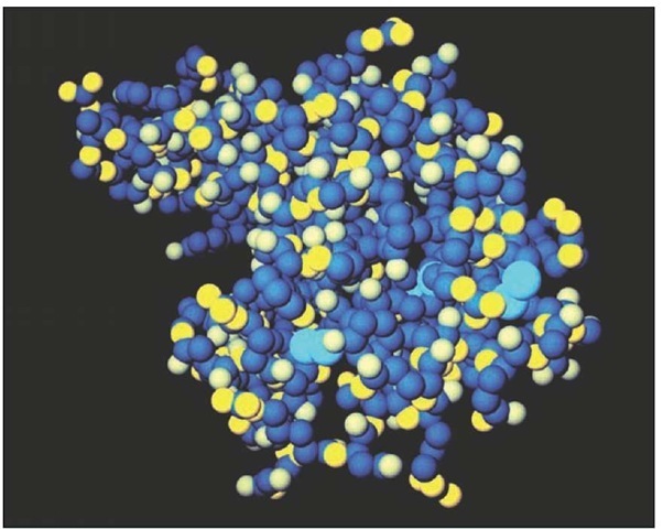
Computer-generated model of lysozyme, an enzyme found in tears and mucus that protects against bacterial infection by literally dissolving the bacteria.
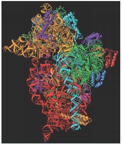
Molecule model of the 30S ribosomal subunit, which consists of protein (corkscrew structures) and RNA (coiled ladders). The overall shape of the molecule is determined by the RNA, which is also responsible for the catalytic function of the ribosome.
DNA is a double-stranded nucleic acid. This macromol-ecule encodes cellular genes and is constructed from adenine, thymine, cytosine, and guanine deoxyribonucleotides. The two DNA strands coil around each other like strands in a piece of rope, creating a double helix. The two strands are complementary throughout the length of the molecule: adenine pairs with thymine, and cytosine pairs with guanine. Thus if the sequence of one strand is known to be ATCGTC, the sequence of the other strand must be TAGCAG.
Phospholipids are the main component in cell membranes; these macromolecules are composed of a polar head group (usually an alcohol), a phosphate, glycerol, and two hydrophobic fatty acid tails. Fat that is stored in the body as an energy reserve has a structure similar to a phospholipid, being composed of three fatty acid chains attached to a molecule of glycerol. The third fatty acid takes the place of the phosphate and head group of a phospholipid.
Polysaccharides are sugar polymers consisting of two or more monosaccharides. Disaccharides (two monosaccharides), and oli-gosaccharides (about three to 12 monosaccharides), are attached to proteins and lipids destined for the cell surface or the extracellular matrix. Polysaccharides, such as glycogen and starch, may contain several hundred monosaccharides, and are stored in cells as an energy reserve.
Basic cellular Functions
There are six basic cellular functions: DNA replication, DNA maintenance, gene expression, power generation, cell division, and cell communication. DNA replication usually occurs in conjunction with cell division, but there are exceptions known as polyploidi-zation (see the Glossary). Gene expression refers to the process whereby the information stored in a gene is used to synthesize RNA or protein. The production of power is accomplished by extracting energy from food molecules and then storing that energy in a form that is readily available to the cell. Cells communicate with their environment and with other cells. The communication hardware consists of a variety of special macromolecules that are embedded in the cell membrane.
DNA Replication
Replication is made possible by the complementarity of the two DNA strands. Since adenine (A) always pairs with thymine (T) and guanine (G) always pairs with cytosine (C), replication enzymes are able to duplicate the molecule by treating each of the original strands as templates for the new strands. For example, if a portion of the template strand reads: ATCGTTGC, the new strand will be TAGCAACG.
DNA replication requires the coordinated effort of a team of enzymes, led by DNA helicase and primase. The helicase separates the two DNA strands at the astonishing rate of 1,000 nucleotides every second. This enzyme gets its name from the fact that it unwinds the DNA helix as it separates the two strands. The enzyme that is directly responsible for reading the template strand, and for synthesizing the new daughter strand, is called DNA polymerase. This enzyme also has an editorial function; it checks the preceding nucleotide to make sure it is correct before it adds a nucleotide to the growing chain. The editor function of this enzyme introduces an interesting problem. How can the polymerase add the very first nucleotide, when it has to check a preceding nucleotide before adding a new one? A special enzyme, called primase, which is attached to the helicase, solves this problem. Primase synthesizes short pieces of RNA that form a DNA-RNA double-stranded region. The RNA becomes a temporary part of the daughter strand, thus priming the DNA polymerase by providing the crucial first nucleotide in the new strand. Once the chromosome is duplicated, DNA repair enzymes, discussed below, remove the RNA primers and replace them with DNA nucleotides.
DNA Maintenance
Every day in a typical human cell, thousands of nucleotides are being damaged by spontaneous chemical events, environmental pollutants, and radiation. In many cases it takes only a single defective nucleotide within the coding region of a gene to produce an inactive, mutant protein. The most common forms of DNA damage are depurination and deamination. Depurination is the loss of a purine base (guanine or adenine), resulting in a gap in the DNA sequence, referred to as a "missing tooth." Deamination converts cytosine to uracil, a base that is normally found only in RNA.
About 5,000 purines are lost from each human cell every day, and over the same time period 100 cytosines are deaminated per cell. Depurination and deamination produce a great deal of damage, and in either case the daughter strand ends up with a missing nucleotide, and possibly a mutated gene, as the DNA replication machinery simply bypasses the uracil or the missing tooth. If left unrepaired, the mutated genes will be passed on to all daughter cells, with catastrophic consequences for the organism as a whole.
DNA damage caused by depurination is repaired by special nuclear proteins that detect the missing tooth, excise about 10 nucleotides on either side of the damage, and then, using the complementary strand as a guide, reconstruct the strand correctly. Deamination is dealt with by a special group of DNA repair enzymes known as base-flippers. These enzymes inspect the DNA one nucleotide at a time. After binding to a nucleotide, a base-flipper breaks the hydrogen bonds holding the nucleotide to its complementary partner. It then performs the maneuver for which it gets its name. Holding onto the nucleotide, it rotates the base a full 180 degrees, inspects it carefully, and, if it detects any damage, cuts the base out and discards it. In this case the base-flipper leaves the final repair to the missing-tooth crew that detects and repairs the gap as described previously. If the nucleotide is normal, the base-flipper rotates it back into place and reseals the hydrogen bonds. Scientists have estimated that these maintenance crews inspect and repair the entire genome of a typical human cell in less than 24 hours.
Gene Expression
Genes encode proteins and several kinds of RNA. Extracting the coded information from DNA requires two sequential processes known as transcription and translation. A gene is said to be expressed when either or both of these processes have been completed. Transcription, catalyzed by the enzyme RNA polymerase, copies one strand of the DNA into a complementary strand of mRNA, which is sent to the cytoplasm, where it joins with a ribosome. Translation is a process that is orchestrated by the ribosomes. These particles synthesize proteins using mRNA and the genetic code as guides. The ribosome can synthesize any protein specified by the mRNA, and the mRNA can be translated many times before it is recycled. Some RNAs, such as ribosomal RNA and transfer RNA, are never translated. Ribosomal RNA (rRNA) is a structural and enzymatic component of ribosomes. Transfer RNA (tRNA), though separate from the ribosome, is part of the translation machinery.
The genetic code provides a way for the translation machinery to interpret the sequence information stored in the DNA molecule and represented by mRNA. DNA is a linear sequence of four different kinds of nucleotides, so the simplest code could be one in which each nucleotide specifies a different amino acid; that is, adenine coding for the amino acid glycine, cytosine for lysine, and so on. The earliest cells may have used this coding system, but it is limited to the construction of proteins consisting of only four different kinds of amino acids. Eventually a more elaborate code evolved in which a combination of three out of the four possible DNA nucleotides, called codons, specifies a single amino acid. With this scheme it is possible to have a unique code for each of the 20 naturally occurring amino acids. For example, the codon AGC specifies the amino acid serine, whereas TGC specifies the amino acid cysteine. Thus a gene may be viewed as a long continuous sequence of codons. However, not all codons specify an amino acid. The sequence TGA signals the end of the gene, and a special codon, ATG, signals the start site, in addition to specifying the amino acid methionine. Consequently, all proteins begin with this amino acid, although it is sometimes removed once construction of the protein is complete. As mentioned above, an average protein may consist of 300 to 400 amino acids; since the codon consists of three nucleotides for each amino acid, a typical gene may be 900 to 1,200 nucleotides long.
Power Generation
Dietary fats, sugars, and proteins, not targeted for growth, storage, or repairs, are converted to ATP by the mitochondria. This process requires a number of metal-binding proteins, called the respiratory chain (also known as the electron transport chain), and a special ion channel-enzyme called ATP synthase. The respiratory chain consists of three major components: NADH dehydrogenase, cytochrome b, and cytochrome oxidase. All of these components are protein complexes with an iron (NADH dehydrogenase, cy-tochrome b) or a copper core (cytochrome oxidase), and together with the ATP synthase are located in the inner membrane of the mitochondria.
The respiratory chain is analogous to an electric cable that transports electricity from a hydroelectric dam to our homes, where it is used to turn on lights or to run stereos. The human body, like that of all animals, generates electricity by processing food molecules through a metabolic pathway called the Krebs cycle, also located within the mitochondria. The electrons (electricity) so generated are transferred to hydrogen ions, which quickly bind to a special nucleotide called nicotinamide adenine dinucleotide (NAD). Binding of the hydrogen ion to NAD is noted by abbreviating the resulting molecule as NADH. The electrons begin their journey down the respiratory chain when NADH binds to NADH dehydrogenase, the first component in the chain. This enzyme does just what its name implies: It removes the hydrogen from NADH, releasing the stored electrons, which are conducted through the chain by the iron and copper as though they were traveling along an electric wire. As the electrons travel from one end of the chain to the other, they energize the synthesis of ATP, which is released from the mitochondria for use by the cell. All electrical circuits must have a ground, that is, the electrons need someplace to go once they have completed the circuit. In the case of the respiratory chain, the ground is oxygen. After passing through cytochrome oxidase, the last component in the chain, the electrons are picked up by oxygen, which combines with hydrogen ions to form water.
The Cell Cycle
Free-living single cells divide as a way of reproducing their kind. Among plants and animals, cells divide as the organism grows from a seed, or an embryo, into a mature individual. This form of cell division, in which the parent cell divides into two identical daughter cells, is called mitosis. A second form of cell division, known as meiosis, is intended for sexual reproduction and occurs exclusively in gonads.
Cell division is part of a grander process known as the cell cycle, which consists of two phases: interphase and M phase (meiosis or mitosis). Interphase is divided into three subphases called Gap 1 (G1), S phase (a period of DNA synthesis) and Gap 2 (G2). The conclusion of interphase, and with it the termination of G2, occurs with division of the cell and a return to G1. Cells may leave the cycle by entering a special phase called G0. Some cells, such as postmitotic neurons in an animal’s brain, remain in G0 for the life of the organism. For most cells, the completion of the cycle, known as the generation time, can take 30 to 60 minutes.
Cells grow continuously during interphase while preparing for the next round of division. Two notable events are the duplication of the spindle (the centrosome and associated microtubules), a structure that is crucial for the movement of the chromosomes during cell division, and the appearance of an enzyme called maturation promoting factor (MPF) at the end of G2. MPF phosphorylates his-tones, proteins that bind to the DNA, and when phosphorylated compact (or condense) the chromosomes in preparation for cell division. MPF is also responsible for the breakdown of the nuclear membrane. When cell division is complete, MPF disappears, allowing the chromosomes to decondense and the nuclear envelope to reform. Completion of a normal cell cycle always involves the division of a cell into two daughter cells, either meiotically or mitotically.
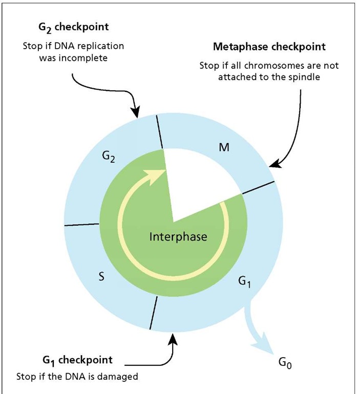
The cell cycle. Many cells spend their time cycling between interphase and M phase (cell division by mitosis or meiosis). Interphase is divided into three subphases: Gap 1(G1), S phase (DNA synthesis), and Gap 2 (G2). Cells may exit the cycle by entering G0. The cell cycle is equipped with three checkpoints to ensure the daughter cells are identical and that there is no genetic damage. The yellow arrow indicates the direction of the cycle.
Cell division is such a complex process that many things can, and do, go wrong. Cell cycle monitors, consisting of a team of enzymes, check to make sure that everything is going well each time a cell divides, and if it is not, those monitors stop the cell from dividing until the problem is corrected. If the damage cannot be repaired, a cell remains stuck in midstream for the remainder of its life. If this happens to a cell in an animal’s body, it is forced to commit suicide, in a process called apoptosis, by other cells in the immediate neighborhood or by the immune system.
The cell cycle includes three checkpoints: The first is a DNA damage checkpoint that occurs in G1. The monitors check for damage that may have occurred as a result of the last cell cycle or were caused by something in the environment, such as UV radiation or toxic chemicals. If damage is detected, DNA synthesis is blocked until it can be repaired. The second checkpoint occurs in G2, where the monitors make sure errors were not introduced when the chromosomes were duplicated during S-phase. The G1 and G2 checkpoints are sometimes referred to collectively as DNA damage checkpoints. The third and final checkpoint occurs in M-phase, to ensure that all of the chromosomes are properly attached to the spindle. This checkpoint is intended to prevent gross abnormalities in the daughter cells with regard to chromosome number. If a chromosome fails to attach to the spindle, one daughter cell will end up with too many chromosomes, while the other will have too few.


