Diabetes
The appearance of life on Earth was made possible to a great extent by the presence of glucose in the oceans and the ability of the first cells to use this sugar as a source of energy. To this day glucose is central to energy metabolism in animals, plants, and microbes. In mammals defects in glucose metabolism and utilization are caused by a disease known as diabetes.
For microbes the process of acquiring glucose and extracting its energy is fairly straightforward. Each cell has receptors that import glucose from the environment, and biochemical pathways that break the sugar down to release the energy it contains. One of the pathways, consisting of a coordinated set of enzymes, is called gly-colysis (meaning sugar splitting), and the other is called the Krebs cycle. These pathways convert the sugar’s energy to ATP, which is used by all cells as an energy source.
Glucose metabolism is more complex in humans and other mammals. In mammals the uptake and utilization of glucose is coordinated by the endocrine system to ensure that the system as a whole has an adequate supply of energy. All cells in an animal’s body have glucose receptors, but cells do not import glucose unless their receptors are bound to a hormone called insulin, which is produced by the pancreas, a large gland located just below the liver. The pancreas has two types of cells, called a (alpha) and i (beta). The a cells produce digestive enzymes that are secreted directly into the large intestine, and the i cells produce insulin. Glucose in the blood stimulates the i cells to make and release insulin; the amount of insulin released is directly proportional to the concentration of glucose in the blood.
One might wonder why the body bothers with such an indirect mechanism: Why not let each cell take up glucose whenever it can? The short answer to this question is that each cell would take up the glucose—a process that requires energy—whether it needed it or not. Dependence on insulin makes it possible for the endocrine system to regulate the uptake of glucose. For example, if the animal has a meal, but each cell already has plenty of ATP on hand, the endocrine system blocks the uptake of glucose everywhere but the liver, which is instructed to convert the glucose into glycogen, a molecule that serves as a storage depot.
Diabetes destroys the i cells’ ability to manufacture insulin, leading to a buildup of glucose in the blood. A chronic elevation of blood glucose levels results in the inappropriate glycosylation (addition of sugar to proteins) of many proteins in the blood, including hemoglobin, the oxygen-carrying protein, as well as many other proteins associated with the cells and tissues. Systemwide protein glycosylation can lead to blindness, heart disease, kidney failure, and neurological disease. Diabetes is a major health problem in North America, where it affects more than 23 million people and causes approximately 500,000 deaths every year. Treatment is very expensive, amounting to about $170 billion annually.
There are two forms of this disease, known as type I and type II diabetes. Type I diabetes is an autoimmune disease, in which the white blood cells attack and destroy the i cells of the pancreas. This form of the disease is sometimes called juvenile diabetes because it occurs predominately in teenagers, although it can strike at any age. Type II diabetes affects older people, usually beginning when they are 50 to 60 years of age. In this case, the disease may be due to a genetic predisposition to short-lived i cells, or it may be due to beta cell burnout brought on by a lifelong preference for a diet that is heavy on sweets. This may account for the fact that more than 90 percent of those suffering from type II diabetes are overweight. At last count, 10 genetic loci were known to be associated with the onset of both types of diabetes.
Osteoporosis
Osteoporosis is a skeletal disorder characterized by weakened bone strength leading to an increased risk of fracture. Healthy bone structure depends on the following elements: mineral content, primarily calcium; the function of osteoblasts, the cells that produce the underlying bone matrix; and osteoclasts, cells that dissolve the bone matrix in preparation for bone remodeling. The steroid hormones estrogen and testosterone control the activity of these cells.
The age-related decline in the amount of these hormones increases the ratio of osteoclasts to osteoblasts and is believed to be the major cause of osteoporosis.
Although bone may seem like an inert material, it is in fact a living tissue like any other tissue or organ in the body. Osteoblasts, the bone-forming cells, are derived from fibroblasts, the principal cellular component of connective tissue. This tissue, consisting of a meshwork of collagen and elastin protein fibers, gives the body shape, holds organs together, and gives skin its elasticity and strength.
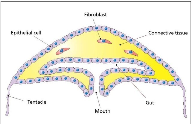
Connective tissue in a jellyfish. The importance of connective tissue is easy to see in a simple animal such as this. The shape of the creature is determined almost entirely by the jellylike connective tissue, which is produced and secreted by the fibroblasts. The internal organs, such as the gut and gonads (not shown), are embedded in the connective tissue. These properties of the connective tissue hold true for all animals, including humans. Note that the size of the epithelial cells, relative to the whole organism, is exaggerated for clarity.
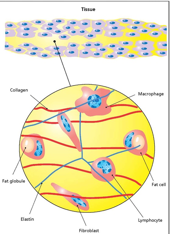
Connective tissue. All of the body’s cells are embedded in connective tissue (colored yellow at the top in a sample of soft, loose tissue). A portion of the connective tissue is shown magnified at the bottom. This tissue consists of collagen fibers, elastin, and three principal cell types: fibroblasts, fat cells, and cells of the immune system (macrophages and lymphocytes).
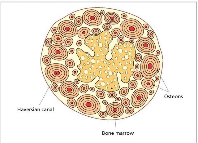
Bone is nothing more than calcified connective tissue. Osteoblasts synthesize bone as a collection of calcified collagen rods, known as osteons, each of which consists of concentric rings. Blood vessels located in the Haversian canals at the center of each osteon supply the tissue with oxygen and nutrients. Every year, as one grows from an infant to an adult, new osteons are added and elongated. This process is so regular that barring traumas or starvation anthropologists have been able to estimate the age of an individual at the time of death by determining the number osteons in the bones.
The human skeleton is subjected to a great deal of stress and strain, which produces many hairline fractures and, in extreme cases, broken bones. Consequently, our bones are in a constant state of repair and remodeling.
Bone structure. Compact or dense bone is constructed from long tubular structures called osteons that are built up in concentric rings. The center of each osteon, called the Haversian canal, contains blood vessels that supply the bone with oxygen and nourishment. The central portion of the bone is porous and is called trabecular bone or, more commonly, bone marrow.
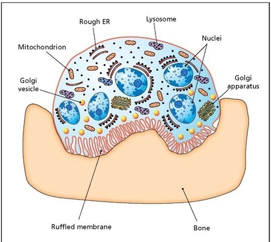
In the absence of fractures or broken bones, remodeling can strengthen specific areas of the skeleton that are subjected to repetitive stress. Estimates of bone replacement in humans range from 10 percent to nearly 20 percent per year, or a complete renewal of the skeleton every five to 10 years. Repair and remodeling are carried out in sequence by the osteoblasts and the osteoclasts. Osteoblasts regulate the process by releasing two growth factors at the appropriate time: macrophage-colony stimulating factor to stimulate the production of osteoclasts, and osteoprotegerin to inhibit osteoclast production. Osteoblasts are large cells with a variable shape that ranges from cuboidal to pyramidal. They have a large nucleus with a single prominent nucleolus and a very extensive endoplasmic reticulum. Osteoclasts are giant cells, about 50 micrometers in diameter, with a remarkable anatomy. These cells have several nuclei, a cytoplasm that is stuffed with organelles, and a cell membrane that is highly ruffled over half of the cell’s surface. The ruffled membrane is brought into contact with the bone, where it secretes acids and hydrolases to dissolve away the bone matrix, after which the cavity is repaired by the osteoblasts. The complete repair cycle takes about 100 days. Osteoclasts function very much like macrophages, phagocytic cells of the immune system, which remove dead or dying cells as well as invading microbes from the tissues. Indeed, osteoclasts and macrophages are both derived from monocytes, a type of stem cell that gives rise to many cells of the immune system.
Bone mineral density (BMD) is a common criterion used to evaluate the onset of osteoporosis, which affects more than 20 million people in North America alone. BMD is usually determined with a special, dual-energy X-ray machine. Women are four times more likely to develop this disease than men. One out of every two women and one in eight men over 50 will have an osteoporosis-related fracture in her or his lifetime. Osteoporosis is caused primarily by hormonal changes that affect women and men as they approach their sixth decade. For women this involves a dramatic drop in estrogen levels at menopause, and for men a reduction in the levels of testosterone at a comparable age.
Osteoclast. The repair and remodeling of bone begins when osteoclasts dissolve away an area of bone by secreting acids and hydrolases, all along their ruffled membrane. Osteoclasts can bore deep into the bone, often forming long tunnels. Once the bone has been removed, osteoblasts move in to repair the area by secreting a new bone matrix.
Osteoporosis is responsible for more than 1.5 million fractures annually, including 300,000 hip fractures, approximately 700,000 vertebral (spinal) fractures, 250,000 wrist fractures, and more than 300,000 fractures at other sites. In the presence of osteoporosis, fractures can occur from normal lifting and bending, as well as from falls. Osteoporotic fractures, particularly vertebral fractures, are usually associated with crippling pain. Hip fractures are by far the most serious and certainly the most debilitating. One in five patients dies one year following an osteoporotic hip fracture. Fifty percent of those people experiencing a hip fracture will be unable to walk without assistance, and about 30 percent will require long-term care.
Current treatments involve calcium and vitamin D supplements (at about 400 to 1,000 IU per day for vitamin D). The preferred calcium source is milk, cheese, or yogurt. Hormone replacement therapy, involving estrogen for women and testosterone for men, has proven to be very effective. Estrogen promotes bone growth by increasing the life span of osteoblasts. It also stimulates osteoblast production of osteoprotegrin (to inhibit osteoclast production) while weakening and killing osteoclasts directly. The effective estrogen dose was once believed to be low enough that cancer induction was not a serious concern, but a major study completed by the National Institutes of Health (NIH) indicated that this may not be so: Estrogen therapy does increase the risk of cancer development. As an alternative, scientists have synthesized a modified estrogen, called estren, which promotes bone growth in mice without simulating cellular growth in ovaries and testes. If these results are confirmed in human clinical trials, it will be possible to treat and prevent osteoporosis in men and women with a single hormone and without fear of cancer induction. Other promising therapies involve a family of drugs known as bisphosphonates (or diphosphonates) and parathyroid hormone.
Bisphosphonates are simple carbon compounds containing two phosphate groups and two variable groups attached to a single carbon atom. These drugs, the best studied of which are risedronate (Actonel) and alendronate (Fosamax), are taken up selectively by the osteoclasts for which they are toxic compounds. As a consequence, the ratio of osteoblasts to osteoclasts increases, and bone remodeling shifts toward bone building. Fosamax increases bone density by 10 percent during the first year and reduces the frequency of fractures by nearly 50 percent during the first three years of treatment. These numbers suggest that bisphosphonates are as effective as estrogen, but the long-term safety (beyond 10 years) of these drugs has not been determined.
Parathyroid hormone (PTH) is a small protein, or peptide, containing 800 amino acids that is normally involved in stimulating the release of calcium from bones and other calcium stores within the body. Thus, it was a surprise when scientists discovered that daily injections of a small amount of this hormone could increase bone density by 10 percent after one year with a 60 percent reduction in the risk of fractures. PTH seems to exert its effect by stimulating the release of insulinlike growth factor-1 (IGF-1) from the liver. IGF-1, in turn, exerts its effect by stimulating the production of osteoblasts. PTH was approved for general use by the U.S. Food and Drug Administration (FDA) in 2002 under the brand name Forteo. The long-term safety of this drug is yet to be determined. In male and female rats, the drug is known to cause osteosarcoma (malignant bone cancer), but currently the incidence of this cancer in patients taking Forteo is unknown.
In addition to drug therapies, regular exercise is recommended as a way to prevent the onset of this disease or to minimize its effects once it has started. A sedentary lifestyle has a devastating effect on bone mass since the induction of osteoblasts (bone-forming cells) is known to be dependent on physical activity. Consequently, a lifelong habit of avoiding exercise is known to be a major risk factor in the onset of osteoporosis.


