Localized Urticaria
Papular urticaria, some of the physical urticarias, and contact urticaria are the entities to consider when urticaria is restricted to a limited area of the body. Dermatographism, cold urticaria, delayed pressure urticaria, solar urticaria, and aqua-genic urticaria are localized wheals produced by specific physical stimuli (i.e., stroking of the skin, cold, sustained pressure, ultraviolet light, and water, respectively).
Papular urticaria Papular urticaria consists of 4 to 8 mm wheals or firm papules, often in grouped clusters and especially on areas of exposed skin. Papular urticaria that is very pru-ritic, persists longer than typical hives, and is located on exposed parts of the body is often caused by insect bites (fleas, bedbugs, scabies, and other mites). The pattern of the eruption corresponds to the biting habits of the offending insect (e.g., mosquito bites often comprise three quasilinear lesions—referred to as breakfast, lunch, and supper), and the seasonal occurrence corresponds to the peak prevalence of that insect. IgE and IgG antibodies against mosquito antigens have been detected in human sera,34 but there have been no reported cases of anaphylaxis or death associated with hypersensitivity to mosquitoes. Arthropod bites are the only known cause of bulla on papular urticaria. Papular urticaria persists for 2 to 10 days and may leave postinflammatory hyperpigmentation. Occasionally, healed lesions may recrudesce when fresh crops appear.
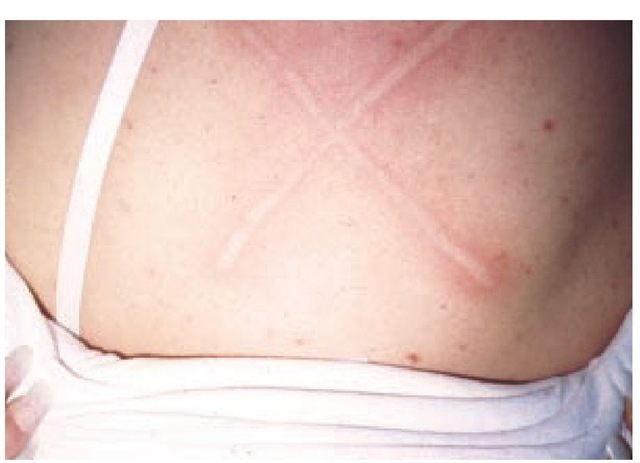
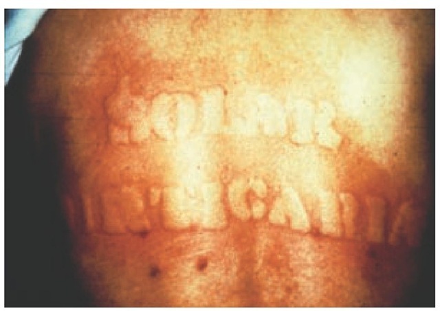
Figure 2 Dermatographism elicited by gentle stroking of the skin of the back.
Dermatographism Firm stroking of the skin may elicit a wheal and erythema in 5% of a healthy population, but only in a minority of these persons does it also cause any pruritus (so-called symptomatic dermatographism) [see Figure 2]. The etiology of dermatographism is uncertain, but passive transfer tests are sometimes positive. Dermatographism (Darier sign) is a common finding in patients with idiopathic urticaria and may be associated with other conditions. For example, dermato-graphism can be elicited in more than 90% of patients with mas-tocytosis.36 Confirmation of mastocytosis always requires biopsy, however. The elicitation of symptomatic dermatographism in patients with urticaria supports the use of both H1 and H2 receptor antagonists, which may more effectively reduce wheal size and duration of urticaria.
Delayed pressure urticaria Urticaria that results from localized, continuous (4 to 6 hours) pressure is seen most often in patients with persistent urticaria without an identifiable cause.38 It may be associated with systemic complaints such as myalgias, arthralgias, and fever. It responds best to aspirin or NSAIDs and poorly to antihistamines.
Cold urticaria The lesions of cold urticaria develop 5 to 30 minutes after exposure to cold and can be caused by wind, bathing, contact, or eating cold foods or drinking cold liquids.39 Although the urticaria may appear during the period of exposure, more often it develops upon rewarming of the skin. The urticaria usually lasts approximately 30 minutes and resolves spontaneously.
Cold urticaria is often idiopathic and acquired. Patients with these lesions usually have a positive response to an ice-cube-challenge test.40 Rare forms of acquired cold urticaria include delayed, localized, and reflex cold urticaria. In delayed cold urticaria, lesions develop several hours after exposure; localized cold urticaria lesions occur only at sites of injections or bites; and reflex cold urticaria lesions present as widespread wheal-ing in response to a fall in core body temperature.
Much rarer than acquired forms of cold urticaria is familial cold urticaria. In this autosomal dominant disorder, lesions appear 30 minutes after exposure to generalized cooling, rather than to local application of cold, and may persist for up to 48 hours.
Solar urticaria Solar urticaria is a rare idiopathic disorder in which erythema heralds a pruritic wheal that appears within 5 minutes after exposure to a specific wavelength of light and dissipates within 15 minutes to 3 hours after onset [see Figure 3].41,42 Solar urticaria is usually provoked by light in the visible spectrum, although the specific wavelength that leads to mast cell degranulation may vary from patient to patient. The severity of the reaction depends on the duration of the exposure, the intensity of the irradiation, and the light spectrum.43 These reactions are believed to result from the development of an anti-genic photoproduct, which then triggers an IgE-mediated response. Patients should usually be referred to an allergist or dermatologist for provocative testing.
Aquagenic urticaria Urticaria that appears 2 to 30 minutes after water immersion, regardless of its temperature or source (seawater or tap water) has been reported in a few patients.44 These pruritic, follicular, cholinergic-like wheals can be reproduced by applying wet compresses to the patient’s back for at least 30 minutes. It is believed that aquagenic urticaria occurs when sensitized mast cells are activated by a water-soluble antigen that diffuses through the epidermis, causing the release of acetylcholine and histamine.
Vibratory urticaria Urticaria that follows massage and vigorous toweling has been described in a single family.
Contact urticaria Immediate contact reactions can appear on normal or eczematous skin within minutes to an hour after exposure. The reaction then will disappear within a few hours. Itching, tingling, or burning accompanied by erythema are the mildest form of contact reactions. They are often caused by cosmetics, fruits, and vegetables. Generalized urticaria after a local contact is a rare phenomenon, but it can occur with some allergens.
Contact reactions may have either immunologic or nonim-munologic mechanisms. Immunologic mechanisms require prior sensitization to the causative agent.
Figure 3 Solar urticaria.
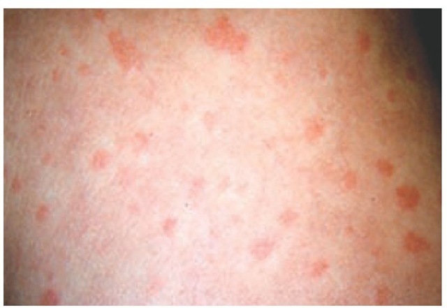
Figure 4 Lesions of generalized urticaria tend to be symmetrical and sometimes have a halo of pallor surrounding the wheal.
The respiratory and gastrointestinal tracts are typically the routes of sensitization, but sensitization to natural latex and some foods may occur through the skin. The substances causing immunologic immediate contact reactions are usually proteins. Foods most commonly involved are fish, shellfish, and wheat flour. Most cases of protein contact dermatitis develop after the person has handled food products for a protracted period. Symptoms usually appear within 30 minutes after direct cutaneous contact with the offending agent.47 Specific IgE antibodies against the causative allergen can be found by skin testing or RAST.
Most immediate contact reactions are nonimmunologic and occur without previous sensitization. These reactions remain localized. The pathophysiology of nonimmunologic immediate contact reactions has not been established, but it may involve direct influence on dermal vessel walls or a non-IgE release of inflammatory mediators. A list of chemicals that cause occupational allergic contact dermatitis can be found on the Internet at http://www.haz-map.com/allergic.htm.
Generalized Urticaria
The clinical features and natural history of generalized urticaria are as varied and unpredictable as the etiology. Generalized lesions tend to be numerous and symmetrical. Characteristically, they are intensely pruritic, especially at onset. Except for cholinergic papular urticaria, little information about the etiology can be obtained from the morphology. Individual lesions fade completely within 24 hours. Occasionally a halo of pallor surrounds the wheal [see Figure 4].
Cholinergic urticaria The lesions of cholinergic urticaria are highly distinctive, consisting of 2 to 3 mm scattered papular wheals surrounded by large, erythematous flares. These lesions are extremely pruritic, and they may affect the entire body but often spare the palms, soles, and axilla.48 Precipitating stimuli include exercise, warm temperature, ingestion of hot or spicy foods, and possibly emotional stress. The condition often remits within several years but can last for more than 30 years. The diagnosis can be made by provocation with exercise or a hot bath. Cholinergic urticaria can be aborted by the prompt application of cold water or ice to the skin, and a refractory period of up to 24 hours can be induced by a hot bath.
Table 4 H1 Antihistamines Available for Treatment of Urticaria
|
Chemical Group |
Agents |
Antihistaminic Activity |
Sedation |
Anticholinergic Activity |
Cost |
|
Ethanolamine derivatives |
Diphenhydramine Clemastine |
+ + |
++/+++ ++ |
++ + |
$ $ |
|
Ethylenediamine derivatives |
Tripelennamine (PBZ) |
++ |
+ |
+ |
$ |
|
Piperidine derivatives |
Azatadine Cyproheptadine Fexofenadine (Allegra)* Loratadine (Claritin)* |
+ ++/+++ |
+ +/++ 0 0 |
+ + 0 0 |
$$ $ $$$ $$$$ |
|
Piperazine derivatives |
Hydroxyzine Cetirizine (Zyrtec) |
+++ +++ |
++ +/0 |
+ 0/+ |
$ $$$$ |
|
Propylamine derivatives |
Acrivastine Brompheniramine Chlorpheniramine Dexchlorpheniramine |
++ + |
0/+ + + + |
0 + + + |
$$$ $ $ $$ |
|
Phenothiazine derivatives |
Promethazine Trimeprazine |
++/+++ ++ |
+++ |
++ ++ |
$ $ |
|
Tricyclic antidepressants |
Doxepin Amitriptyline |
+++++ +++++ |
+++ +++/++++ |
+++ +++ |
$ $ |
"Considered second-generation antihistamines, which are nonsedating (Zyrtec less so) and have other anti-inflammatory properties besides being antihistaminic.
Physical Examination
Recognition of urticaria does not usually present a problem. Unfortunately, except for the contact and physical urticarias, the examination does not facilitate identification of the cause. Episodes of angioedema occur in half the patients presenting with persistent urticaria. The individual swellings of angioede-ma always last longer than an individual hive and are almost always nonpruritic.
Laboratory Evaluation
The use of laboratory tests in patients with urticaria should be directed toward confirmation of diagnoses suggested by the history and physical examination. Routine laboratory testing should not be performed, because it has consistently proved disappointing for the identification of an etiology. A skin biopsy is indicated if the diagnosis of urticaria is in question. A biopsy should be performed on any urticaria that lasts more than 24 hours, is only mildly pruritic or nonpruritic, is associated with vesicles or bullae, or does not respond to appropriate therapy. The subtleties of the histologic variances demand interpretation by a dermatopathologist.
Treatment
Eliminating or avoiding the triggers of mast cell activation is the basis of treatment for urticaria. However, this strategy may be impractical in patients with persistent idiopathic urticaria, which usually has multiple triggers. Any underlying disease should be treated. Idiopathic urticaria is managed symptomati-cally. Fortunately, the hyperreactive state in patients with idio-pathic urticaria eventually resolves spontaneously.
H1 Receptor Antagonists
When used appropriately, antihistamines can offer significant relief to most patients with urticaria. The more the skin lesions resemble the triple response of Lewis, the better they respond to antihistamine treatment. Urticarial vasculitis and delayed pressure urticaria are resistant to antihistamines. Antihis-tamines compete with histamine for H1 receptor sites on effector cells and thereby prevent, but do not reverse, responses mediated by histamine alone. There are eight recognized chemical groups of H1 receptor antihistamines; all effectively compete for H1 receptor sites [see Table 4]. Among these groups are tri-cyclic antidepressants, which also have potent antihistaminic activity.
The choice of antihistamine is based on its effectiveness, frequency of administration, and side-effect profile. The dose of the agent selected should be increased to tolerance; if adequate relief is not achieved at the maximal tolerated dose, a drug from another group can be added. Patients do not all respond in the same way to agents from each group. Most of the so-called first-generation (sedative) antihistamines are virtually equivalent in effectiveness, with the major differences being the degree of sedation or anticholinergic effects. Activation of H1 receptors in the brain is responsible for alertness; inhibiting these sites with antihistamines results in sedation. Second-generation (nonsedating) antihistamines tend not to cause drowsiness, because they cross the blood-brain barrier poorly.49 Many patients find the itching and urticaria to be most troublesome in the evening and at night, so a useful strategy is to combine sedating antihistamines given at bedtime with nonsedating an-tihistamines during the day. This combination is effective, promotes compliance, and is economical. Tachyphylaxis has not been noted with H1 receptor antagonists.
Because other mediators besides histamine are involved in urticaria, antihistamines are not a panacea. Also, none of the antihistamines have the ability to displace histamine from the H1 receptor site, so the best clinical results are attained when the antihistamines occupy those receptors before the arrival of histamine; hence, round-the-clock dosing is necessary for patients with persistent symptoms.
An effective cocktail for persistent urticaria is fexofenadine (180 mg) or loratidine (10 mg) in the early morning and ceti-rizine (10 to 20 mg) in the early evening. If this is insufficient, the tricyclic antidepressant doxepin, 10 to 50 mg, can be added at bedtime. (A single dose of doxepin suppresses the histamine-in-duced wheal and flare for 4 to 6 days.50) This cocktail controls symptoms in more than three quarters of patients with persistent urticaria. Prednisone, 0.5 to 1.0 mg/kg/day, should be used only for patients with refractory idiopathic urticaria or with ur-ticarial vasculitis. The goal of treatment should not be to attain a hive-free status but, rather, to minimize compromise of the patient’s quality of life from both the disease and its treatment.
In urticaria with an identifiable cause, antihistamines are discontinued once the substance is gone from the body. In persistent urticaria, antihistamines can be sequentially discontinued when patients have been completely free of hives for at least 96 hours. At that point, the morning dose of nonsedating antihistamines can be discontinued. If the patient is still symptom free after another 96 hours, the doxepin dosage can begin to be reduced and, lastly, the cetirizine can be discontinued.
H2 Receptor Antagonists
Human skin has H2 receptors as well as H1 receptors. H2 receptors are present on the cutaneous arterioles, and their activation can result in vasodilatation (noted as flushing). For that reason, combining H2 antagonists with H1 antagonists can be helpful in patients who have prominent flushing, der-matographism, or angioedema.51 The available evidence does not justify the routine addition of H2 antagonists to H1 antagonists in patients with persistent urticaria or urticarial vasculitis.


