Human and Animal Herpesviruses
The herpes group of viruses is composed of at least eight human viruses and numerous animal viruses [see 7:XXX1 Viral Zoonoses]. The human herpesviruses include herpes simplex virus types 1 and 2 (HSV-1 and HSV-2), varicella-zoster virus (VZV), cytomegalovirus (CMV), Epstein-Barr virus (EBV), and human herpesvirus types 6 (HHV-6), 7 (HHV-7), and 8 (HHV-8, also known as Kaposi sarcoma-associated herpesvirus [KSHV]).
All human herpesviruses are of similar size and morphology and are characterized by a core, 30 to 60 nm in diameter, that contains a double-stranded DNA genome; a nucleocapsid, 95 to 100 nm, that exhibits icosahedral symmetry; and a lipoprotein envelope with glycoprotein projections that has a diameter of 120 to 250 nm. Herpesviruses replicate primarily in cell nuclei, and a protein envelope is added as the virus passes through the nuclear membrane.
Human herpesviruses share the properties of latency and reactivation. Members of the group can cause productive lytic infections, in which infectious virus is produced and cells are killed, or nonproductive lytic infections, in which viral DNA persists but complete replication does not occur and cells survive. After acute lytic infections, herpesviruses often persist in a latent form for years; periodic reactivations are followed by recurrent lytic infections. Sites of latency vary: HSV and VZV persist in neural ganglion cells, EBV persists in B cells, and CMV probably remains latent in many cell types. The sites of latency for HHV-6 and HHV-7 have not been identified, although both herpesviruses have been detected in salivary glands.
All human herpesviruses have a worldwide distribution [see Sidebar Herpesvirus Information on the Internet]. Considerable efforts are being directed toward the development of vaccines and antiviral agents that will be active against herpesviruses.
Herpes Simplex Virus
HSV-1 and HSV-2 can be distinguished by a variety of properties, including clinical and epidemiologic patterns, antigenici-ty, DNA base composition, biologic characteristics, and sensitivity to various physical and chemical stresses.1 Advances in molecular biology technology have proved that HSV-1 and HSV-2 share certain antigens (e.g., glycoprotein B) but differ with respect to other antigens (e.g., glycoprotein G). Restriction enzyme analysis of HSV DNA and other molecular techniques are used to identify individual isolates.
EPIDEMIOLoGY and ETIOlOGY
Humans are the only known natural hosts for HSV, although animals can readily be infected experimentally. HSV-1 primary infection occurs mainly in childhood, whereas HSV-2 infection occurs predominantly in sexually active adolescents and young adults. The prevalence of HSV-2-specific antibodies in the United States increases from less than 6% in those younger than 19 years to more than 25% in those older than 30 years.2 In older age groups, changes in prevalence are negligible. Independent predictors of HSV-2 seropositivity include female gender, black race, increasing age, less education, more lifetime sex partners, prior occurrence of syphilis or gonorrhea, and lack of HSV-1 an-tibody.3 In the United States, changes in sexual mores resulted in an age-adjusted seroprevalence of HSV-2 infection in the 1990s that was 30% higher than the seroprevalence in the 1970s; HSV-2 is now detectable in one of five persons older than 12 years.2 Approximately one third of new HSV-2 infections are sympto-matic.4 Prevalence of antibody to HSV-1 in different populations ranges from 44% in persons 12 to 19 years of age to above 80% in those older than 60 years.2 Between 10% and 25% of adults are dually infected with HSV-1 and HSV-2.
Direct contact with infected secretions is the principal mode of transmission. HSV-1 is usually transmitted by an oral route and HSV-2 by a genital route. Although virus titers are higher and transmission is more likely when lesions are present, asymptomatic excretion of the virus is common.
| Herpesvirus Information on the Internet |
| Generalhttp://www.nesfile.com/xlp.htm
Herpes Viruses Weekly http://www.cdc.gov/std/treatment/TOC2002TG.htm |
| Sexually Transmitted Disease Treatment Guidelines http://www.cdc.gov/mmwr/preview/mmwrhtml/00021095.htmRecommended infection-control practices for dentistry |
| http://www.cdc.gov/mmwr/preview/mmwrhtml/00001053.htmPerspectives in disease prevention and health promotion: condoms for prevention of sexually transmitted diseases |
| HSVhttp://www.cdc.gov/std/Herpes/STDFact-Herpes.htm
Patient information on genital herpes |
| http://www.cdc.gov/mmwr/preview/mmwrhtml/00047449.htmCase definitions for infectious conditions under public health surveillance |
| VZVhttp://www.cdc.gov/nip/diseases/varicella/vac-chart.htm |
| Varicella patient information in brief |
| http://www.cdc.gov/nip/diseases/varicellaFrequently asked questions about varicella and varicella vaccine |
| http://www.cdc.gov/od/oc/media/fact/chickenp.htm |
| Facts about chickenpox (varicella) from the CDC Office of Communications, Division of Media Relationshttp://www.cdc.gov/epo/mmwr/preview/mmwrhtml/00022690.htm |
| ACIP recommendation: varicella-zoster immune globulin for the prevention of chickenpoxhttp://www.cdc.gov/mmwr/preview/mmwrhtml/00042990.htm |
| Recommendations and Reports—June 27, 1996/Vol. 45/No. RR-1-Preview (The first statement by the Advisory Committee on Immunization Practices [ACIP] on the use of live, attenuated varicella virus vaccine) |
| CMVhttp://www.cdc.gov/ncidod/diseases/cmv.htm
Patient information on cytomegalovirus http://www.cdc.gov/hiv/pubs/brochure/oi_cmv.htm Division of HIV/AIDS Prevention Brochures: You Can Prevent CMV |
| Herpesvirus Simiaehttp://www.cdc.gov/epo/mmwr/preview/mmwrhtml/00015936.htm
Guidelines for prevention of herpesvirus simiae (B virus) infection |
HSV-2 shedding from the genital tract can occur in seropositive persons who have no history of genital HSV infection.5 Thus, transmission occurs frequently, even in the absence of lesions. HSV-2 is transmitted more efficiently from males to females than from females to males. Au-toinoculation to other skin sites also occurs, more often with HSV-2 than with HSV-1. Spread of infection through contact with oral secretions may be an occupational hazard for respiratory care and dental care providers; thus, gloves should be worn when fingers are placed in patients’ mouths. Fomites, including toilet seats and towels, are not important modes of transmission. Recurrences are frequent with both HSV-1 and HSV-2 infections, usually as a result of endogenous reactivation. In the United States, lip or perioral recurrences develop in 20% to 40% of the population. Precipitating factors are sunlight, wind, local trauma, fever, menstruation, and emotional stress. Ocular herpes is present in about 5% of all patients seen at ophthalmology clinics; 25% to 50% of ocular HSV infections recur within 2 years. Of all the primary cases of genital herpes in the United States each year, 60% to 80% will recur. Although most genital recurrences represent reactivation, exogenous reinfection can also occur. Clinically significant recurrences tend to decrease over time.6
Pathogenesis
After the initial replication of the virus in epithelial cells, cytol-ysis and local inflammatory reactions develop, resulting in the characteristic lesion—a superficial vesicle on an inflammatory base. Multinucleated cells and Cowdry type A inclusion bodies are present. Subsequent lymphatic spread to regional nodes and viremic spread to other organs may occur, depending on the immune competence of the host. Viremia can be demonstrated in malnourished children, in certain adults with depressed T cell-mediated immunity, and, occasionally, in immunocompe-tent persons.
After initial infection, HSV travels along sensory nerve pathways to ganglion cells, the site of latent infection. The viral DNA persists, only to become reactivated by certain stresses. After reactivation, the virus reverses its course and spreads peripherally by sensory nerve pathways. Once HSV reaches cutaneous sites, cell-to-cell spread occurs until host immune mechanisms limit further dissemination. Various mechanisms are engaged in host responses to HSV, including T cell and natural killer cell cytotox-icity, macrophage activation, production of antibody, and production of interferon.
Clinical Syndromes
Oral-Labial Herpes
In patients younger than 5 years, primary HSV-1 infection is most often asymptomatic; when symptomatic, it presents as gin-givostomatitis or pharyngitis. After an incubation period of 2 to 12 days, fever and sore throat develop. Small vesicles are observed on the oral mucosa and pharynx. Mouth pain may be severe, breath is fetid, and cervical adenopathy is present. In adolescents and young adults, posterior pharyngitis and tonsillitis may be the primary problem. The differential diagnosis includes streptococcal pharyngitis, aphthous stomatitis, Stevens-Johnson syndrome, herpangina, and infectious mononucleosis. Symptoms and signs often persist for 10 to 21 days, and autoinocula-tion to other sites, such as fingers and eyes, is common. Oral shedding may persist for several weeks (mean, 7 to 10 days).
Recurrent herpes labialis is a shorter and milder affliction, often heralded by local pain or tingling for a few hours. HSV-1 oral-labial lesions recur more often than HSV-2 oral-labial le-sions.3 Vesicles appear most often on the vermilion border and are painful. The lesion is usually small (< 1 cm2), and the progression from vesicle to ulcer to crust is rapid (< 96 hours). Healing is complete within 8 to 10 days. Systemic complaints are uncommon in recurrent herpes labialis.
Ocular Herpes
Most ocular herpetic infections are caused by HSV-1. Primary infections may present as unilateral follicular conjunctivitis, blepharitis, or corneal epithelial opacities. Healing is usually complete within 2 to 3 weeks. Recurrences may take the form of keratitis (more than 90% of cases are unilateral), blepharitis, or keratocon-junctivitis. Branching dendritic ulcers, usually detected by fluo-rescein staining, are virtually diagnostic and are often associated with diminished visual acuity. Deep stromal involvement may result in scarring, corneal thinning, and abnormal vasculariza-tion, with resultant blindness or rupture of the globe.
Genital Herpes
HSV-2 is the causative agent in 70% to 95% of primary genital herpesvirus infections. After an incubation period of 2 to 7 days, fever, malaise, and inguinal adenopathy develop; these symptoms are associated with the appearance of vesicular lesions. In men, lesions are often on the glans penis or penile shaft [see Figure 1]; in women, lesions may involve the vulva, perineum, buttocks, cervix, or vagina. The differential diagnosis includes syphilis, chancroid, Behcet syndrome, erythema multiforme, and can-didiasis. In women, lesions rapidly ulcerate and become covered with exudate, with resultant vaginal discharge. Urethral involvement sometimes results in dysuria or urinary retention. Signs and symptoms of primary genital herpes often persist for several weeks before complete healing. Previous infection with HSV-1 may reduce the severity and duration of a first episode of genital herpes caused by HSV-2.
Extragenital lesions develop during the course of primary infection in 10% to 18% of patients. Aseptic meningitis is not uncommon during primary genital herpes, particularly in women, and in rare instances, herpetic sacral radiculomyelitis occurs.
Images removed due to full nudity regulations.
Figure 1 Herpes simplex virus genital lesions in men often present as grouped vesicles on the penile shaft.
Figure 2 Perianal herpes simplex infections in patients with compromised immunity may be severe and prolonged.
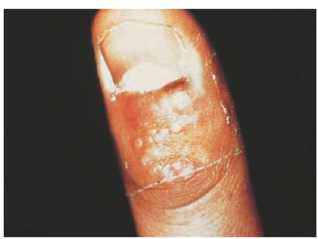
Figure 3 Herpetic whitlows, which occur on fingers, are often misdiagnosed as staphylococcal infections.
Urinary retention may occasionally complicate primary genital herpes, particularly in women.
Recurrent episodes of genital herpes are usually shorter and milder than primary episodes but still affect women more severely than men. Genital HSV-2 infections recur more often than genital HSV-1 infections, and on rare occasions, the two infections can be found simultaneously in lesions.7,8 Moreover, patients dually infected with HSV-1 and HSV-2 have fewer recurrences of genital herpes than those infected with HSV-2 alone.2 With either virus type, after a variable prodrome of tenderness, itching, or tingling, lesions develop on the penis, labia minora, labia majora, perineum, mons pubis, or buttocks. Healing occurs in 6 to 10 days and is usually uncomplicated; frequent asymptomatic shedding occurs, however, particularly in women.5,9 HSV-2 may also cause benign, recurrent lymphocytic meningitis that may be associated with recurrent genital lesions.
Perianal and Anal Herpes
Perianal and anal HSV-2 infection is an important problem in men who have sex with men. Pain, itching, tenesmus, discharge, fever, chills, sacral paresthesias, headache, and difficulty in urinating may all occur. Vesicles and ulcerations may lead to an ery-thematous cryptitis with inguinal adenopathy. Herpes proctitis is often prolonged and severe in patients with AIDS [see Figure 2].
Other Herpes Syndromes
Herpetic whitlow Primary finger infections, or whitlows, usually involve one digit and are characterized by intense itching or pain followed by the formation of deep vesicles that may coalesce [see Figure 3]. Among the general public, whitlows are most often caused by HSV-2, whereas among medical and dental personnel, HSV-1 is the principal culprit. The lesions gradually resolve in 2 to 3 weeks, unless they are mistakenly incised, in which case healing may be delayed by secondary bacterial infection. Recurrent whitlows commonly appear and are sometimes associated with severe local neuralgia.
Neurologic complication Encephalitis, a severe form of HSV infection, is discussed elsewhere [see 11:XVI Acute Viral Central Nervous System Diseases]. HSV-1 has also been implicated as an etiologic agent in Bell palsy11 and in rare cases of recurrent self-limited meningitis (Mollaret meningitis).12
Infection in the immunocompromised host Disorders of T cell-mediated immunity are associated with more severe HSV infections. In clinical settings such as organ transplantation, lym-phoreticular neoplasm, or AIDS, HSV infection is often slow to heal and may disseminate cutaneously or to visceral organs. Certain skin conditions, such as eczema and burns, are associated with cutaneous but not visceral dissemination. In rare instances, HSV infection during pregnancy or in the elderly is complicated by visceral dissemination, particularly to the liver. Intubation or catheterization of debilitated patients may facilitate the spread of infection; for instance, herpes esophagitis often complicates long-term use of nasogastric tubes.
Neonatal infection Between one in 2,500 and one in 10,000 births are complicated by HSV infection, usually HSV-2. HSV neonatal infection can be localized or disseminated and results from transmission of the virus to the infant at the time of delivery, either by ascending infection after premature membrane rupture or by passage of the infant through an infected genital tract. The risk of transmission is increased in premature births, after prolonged membrane rupture, and with the use of fetal scalp monitor electrodes. About 40% to 50% of infants born to mothers with primary infections are at risk for the development of severe disease, whereas fewer than 8% of those born to women with recurrent herpes are at risk for severe disease.1,13 Maternal infection is often asymptomatic at the time of delivery, and asymptomatic shedding may occur in women with no known history of genital herpes. In women with a history of genital HSV infection, genital HSV can be detected at delivery in approximately 2% by use of tissue culture and in 14% by use of polymerase chain reaction (PCR) techniques.
Infection becomes apparent several days to weeks after delivery. Newborns often present with vesicles or conjunctivitis, or a syndrome resembling neonatal sepsis may be evident. Neurologic signs such as seizures, cranial nerve palsies, and lethargy often predominate and are accompanied by cerebrospinal fluid pleocytosis. Disseminated infection may involve the liver, lungs, or adrenal glands. If untreated, disseminated or central nervous system infection is fatal in more than 70% of patients, whereas localized disease is generally self-limited. Treatment has greatly reduced the mortality from severe infection [see 11:XVI Acute Viral Central Nervous System Diseases].
Diagnosis
A variety of tissue culture systems support the replication of HSV, and virus isolation from specimens collected early in the course of infection is the diagnostic method of choice. The characteristic cytopathic effect of the virus is often detectable within a period of 24 to 48 hours. Typing of isolates can be accomplished most readily by immunofluorescence with monoclonal antibodies directed against type-specific antigens. Scrapings or tissue specimens can sometimes be tested directly for herpesvirus antigens by immunofluorescence or immunohistochemistry. Alternatively, scrapings may be prepared by Giemsa or Wright stain and examined for the presence of multinucleated giant cells, which indicates infection with HSV or VZV. Serologic techniques that accurately differentiate HSV-1 from HSV-2 infections are now commercially available. Such tests can be used to confirm a diagnosis of primary HSV infection, but they are seldom helpful in diagnosing recurrences. Serologic techniques can also establish a diagnosis in patients with atypical complaints, identify asymptomatic carriers, and identify persons at risk.15 PCR detection of HSV DNA in CSF has become the standard means of diagnosing HSV encephalitis. For patients with HSV encephalitis, PCR results are often positive within 24 hours of the onset of symptoms, and test results may remain positive during the first week of illness.16
Prevention
No HSV vaccine has been approved for general use, although preliminary studies of glycoprotein-D-adjuvant vaccines suggest efficacy in certain populations (i.e., women seronegative for both HSV-1 and HSV-2) but not others.17 Prophylactic measures that prevent contact with the virus may help in avoiding primary infection. For instance, the use of condoms may prevent sexual transmission when either sexual partner has a history of genital HSV infection. Application of sunscreens to susceptible skin areas before exposure to ultraviolet light can prevent reactivation of HSV. Medical and dental personnel who treat HSV-positive patients should wear gloves to prevent contact with infected areas.
The best strategy for the prevention of neonatal herpes may be close physical examination at the time of labor.18 Cesarean section is indicated to prevent perinatal infection when lesions consistent with the diagnosis of genital herpes are noted during labor. If primary genital herpes is detected during the third trimester of pregnancy, acyclovir in conventional doses should be considered for the mother during the peripartum period and for the newborn post partum [see Treatment, below]. Acyclovir in late pregnancy may also be useful in women with recurrent genital HSV infections; placebo-controlled trials have shown that 400 mg three times daily reduced lesions and HSV excretion at delivery.

