Deep Tissue Infections
Infections originating in the pharynx may extend by contiguous spread to the deep tissues of the pharynx and neck. In the neck, numerous fascial planes create a variety of potential spaces where infection can become loculated to form a phlegmon or a full-fledged abscess. Although these processes are now uncommon, prompt recognition is mandatory because antibiotics and surgical drainage are required to control infection and to prevent obstruction of the airway, invasion of vital neurologic and vascular structures, and spread of infection to the mediastinum and bloodstream.
Peritonsillar abscess
Peritonsillar abscess, also called quinsy throat, is a complication of streptococcal tonsillitis most often seen in adolescents and young adults. Group A streptococci are the primary cause of the condition, although most peritonsillar abscesses also harbor mixed oral bacteria, with a predominance of anaerobes. Patients have fever and sore throat, often with pain referred to the ear. Dysphagia prevents the patient from swallowing saliva, commonly causing drooling; edema and pain produce a characteristic muffled, so-called hot-potato voice. The affected tonsil is visibly displaced forward, downward, and toward the midline; the soft palate may be edematous. Trismus occurs in some patients. CT scans are helpful in diagnosis and management. Treatment consists of parenteral penicillin and surgery. The traditional approach consists of immediate incision and drainage, followed by tonsillectomy 4 to 6 weeks later; early tonsillectomy has also produced excellent results. Treatment with needle aspiration and oral antibiotics has also been successful; 80% to 92% of patients with peritonsillar abscess can be cured with this approach, thereby obviating hospitalization and surgery.
Retropharyngeal and parapharyngeal infections
Retropharyngeal infections are most common in childhood because the lymph nodes in this region atrophy during adult life. Patients have fever and systemic toxicity, as well as neck pain, dysphagia, muffled voice, and respiratory stridor. Physical findings include erythema and bulging of the posterior wall of the pharynx. CT scans and lateral-view x-rays of the neck are extremely useful, invariably demonstrating soft tissue swelling and forward displacement of the larynx [see Figure 3]. Although the bacterial agent of retropharyngeal infections is not determined in many patients, streptococci and other mouth flora predominate. Penicillin is the traditional antibiotic of choice, but agents that provide a broader spectrum of antibacterial coverage may be justified until culture data are available. Surgical drainage is vital to prevent asphyxiation and extension of infection to the mediastinum. Infections of the retropharyngeal space must be distinguished from infections of the prevertebral space.
The prevertebral space lies posterior to the retropharyngeal space and is separated from it by the prevertebral fascia. Infection of the prevertebral space often originates from osteomyelitis of the cervical vertebrae. This infection is frequently caused by staphylococci and may lead to spinal cord damage unless treated through an approach that combines immobilization, antistaphylococcal antibiotics, and external drainage.
The parapharyngeal space is demarcated by the parotid gland and the internal pterygoid muscle laterally and by the superior constrictor muscle medially. The internal jugular vein, carotid artery, and cranial nerves IX, X, and XII pass through the parapharyngeal space. Infection can reach the parapharyngeal space from the pharynx or from parotid or dental foci. Patients have severe trismus, externally visible inflammation behind the angle of the jaw, and inflammation in the lateral wall of the pharynx, with medial displacement of the tonsil. Treatment consists of intravenous penicillin and drainage; CT-guided needle aspiration may be effective in some patients, but surgical drainage from behind the angle of the jaw is required in others.
Infections of the parapharyngeal space occasionally spread to the jugular vein and cause the syndrome of postanginal sepsis (Lemierre syndrome), which is characterized by the presence of septic phlebitis, septic pulmonary emboli, and anaerobic bacteremia. Pharyngitis and dental infections may also lead to postanginal sepsis. Facial swelling is an early diagnostic clue to this syndrome. CT is important for detecting abscesses, and ultrasonography is helpful for identifying jugular vein thrombosis. Anaerobic gram-negative bacilli, such as Fusobacterium species, are typically responsible for the condition. Management requires intravenous administration of penicillin; metro-nidazole and clindamycin are useful alternatives. Heparin, venous ligation, and surgical thrombectomy have also been advocated, but data do not allow firm guidelines to be established for these interventions.
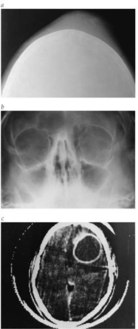
Figure 3 (a) An oblique-view x-ray of the skull of a 16-year-old boy with fever, headache, and doughy edema of the forehead shows soft tissue swelling and a small area of underlying cortical irregularity indicative of osteomyelitis. (b) Sinus film reveals severe pansinusitis. (c) Because of subtle personality changes, a CT scan was taken; it revealed a large frontal lobe abscess. At surgery, 45 ml of pus was evacuated. Anaerobic streptococci were isolated from the subperiosteal collection and the brain abscess. Prolonged penicillin therapy and surgical management led to recovery.
Ludwig angina
Ludwig angina is a cellulitis of the submandibular, sublin-gual, and submental regions. In 86% of patients, the infection originates from a dental focus. Clinical features include fever, marked toxicity, and a rapidly progressive brawny edema in the floor of the mouth and the anterior neck. Elevation of the tongue impedes swallowing, and airway obstruction may be lethal. Streptococci and mouth flora are the most common etio-logic agents, but H. influenzae, staphylococci, and gram-negative bacilli have also been implicated; broad antibiotic coverage may be necessary initially. Because endotracheal intubation can provoke laryngeal spasm, tracheostomy may be necessary to preserve the airway. Surgical decompression may be required.
Acute Epiglottitis (Supraglottitis)
No infection of the upper respiratory tract is more rapidly progressive or more lethal than acute epiglottitis, also known as supraglottitis. Acute epiglottitis occurs most commonly in children between 2 and 8 years of age and is more frequent in boys. The incidence of epiglottitis in childhood is declining rapidly in populations that have received H. influenzae type b vaccinations. Cases in adults appear to be increasing, however, perhaps because of improved diagnosis.
Etiology
The major cause of acute epiglottitis in children and adults is H. influenzae type b. Other pathogens, including pneumococci, streptococci, staphylococci, and Klebsiella pneumoniae, can produce an identical syndrome. Although viral epiglottitis is rare, this syndrome can occur secondary to infection with herpes simplex virus type 1.51 In the immunocompromised host, additional organisms may be responsible, including Candida and Kingella kingae; fortunately, such cases are rare.
Diagnosis
Epiglottitis begins with startling rapidity; severe sore throat and fever progress rapidly to dysphagia, with retention of secretions and drooling. Systemic toxicity is marked; if the illness is not treated, dyspnea and progressive respiratory obstruction occur in a matter of hours.
Although uncommon, acute epiglottitis in adults often extends to the supraglottic structures, and edema may be more prominent than acute inflammation. Whereas children with acute epiglottitis usually present with respiratory distress, adults may first complain of pharyngeal symptoms (severe pain and odynophagia) before developing respiratory symp-toms.52 The course of the disease in adults is slower than it is in children, often extending over several days because of the larger diameter of the adult airway.
Acute epiglottitis is a medical emergency. The key to the diagnosis is a swollen, edematous, cherry-red epiglottis. Simple inspection of the pharynx is usually unrewarding. Furthermore, any instrumentation, even a tongue blade, can provoke spasm and total airway obstruction, although adults are at lower risk for this complication. Therefore, unless acute respiratory distress is present, a lateral-view x-ray of the neck should be taken immediately. If the film does not demonstrate epiglottal edema, indirect laryngoscopy can be undertaken; if edema is present, however, the diagnosis is confirmed, and instrumentation is unnecessary. In all cases, patients must be monitored continuously for signs of respiratory obstruction.
Differential diagnosis
In the differential diagnosis in children, it is important to distinguish between epiglottitis and croup caused by viral laryngitis with laryngeal and subglottal edema and airway obstruction. Croup usually affects younger patients and progresses more slowly than epiglottitis, and patients with croup present with hoarseness and a characteristic cough. In addition, in croup, the epiglottis is normal or only minimally inflamed on physical or radiographic examination. Cold, humidified oxygen is the mainstay of treatment of croup, and close observation is vital because emergency intubation or tracheostomy may be necessary. In children, bacterial tracheitis or membranous croup may present as severe laryngotracheitis, requiring antibiotic therapy and respiratory support. In patients of all ages, angioneurotic edema of the larynx, foreign bodies lodged in the upper airway, or impingement on the airway by various mass lesions may initially be confused with epiglottitis, but these conditions should be readily recognizable on physical examination and radiography.
Treatment
Although tracheostomy is the traditional means of securing a patent airway in patients with epiglottitis, nasotracheal intubation is safe and effective. As a result, direct laryngoscopy by an experienced observer who is prepared to proceed with intubation (or, if necessary, tracheostomy) can be relied on for diagnosis when taking x-rays would produce excessive delay.
Because there is no margin for error, initial therapy with high-dose intravenous antibiotics effective against H. influenzae (including penicillinase-producing strains), other gram-negative bacilli, and staphylococci is warranted. Many regimens are available, including cefuroxime or cefamandole, ampicillin-sul-bactam, meropenem, imipenem-cilastatin, and a third-generation cephalosporin combined with a penicillinase-resistant penicillin. The choice of drugs depends on the results of throat and blood cultures and on ampicillin-sensitivity testing of H. influen-zae isolates. A mist tent may be helpful. Steroids are sometimes advocated to reduce the edema, but their effectiveness has not been tested in controlled clinical trials. Above all, preservation of the airway is critical; nasotracheal intubation or tracheostomy is required in about half of all cases. If the airway can be protected while antibiotics take effect, most patients can be saved.
Miscellaneous Upper Respiratory Tract Infections
Many other infectious processes can invade the upper respiratory tract and adjacent regions of the head and neck. Common viral infections of the oropharynx are primary herpes simplex gingivostomatitis and adenovirus and coxsackievirus infections. Spirochetes play a role in ulcerative gingivitis and periodontitis. Actinomyces israelii can produce chronic burrowing infections of the gums and jaw, occasionally with secondary involvement of the respiratory tract. Although rare in the United States, rhino-scleroma, believed to be caused by Klebsiella rhinoscleromatis, can occur in immigrants from developing countries or in HIV-infect-ed persons. S. aureus may produce acute bacterial sialadenitis or suppurative parotitis; less commonly, gram-negative bacilli or even anaerobes produce parotitis in hospitalized patients.
Many bacterial species have been implicated in a variety of dental infections. Mixed infections with normal mouth flora can occur in debilitated patients. Fusobacteria and spirochetes can cause gingivitis (trench mouth) or necrotic tonsillar ulcers (Vincent angina); patients have foul breath, pain, and dirty-gray membranous inflammation that bleeds easily. Oral irrigations and penicillin are effective therapy. A similar combination of spirochetes and other bacteria can produce a rare but extremely serious invasive gangrene of the mouth, cancrum oris, which occurs only in malnourished infants or in patients with advanced malignant disease or immunosuppression. Anaerobes may play a role in chronic or recurrent tonsillitis.38 Dental infection is the leading cause of necrotizing cervical fasciitis.
Many noninfectious processes in these regions can produce symptoms that mimic bacterial infection. Aphthous stomatitis may be the most common example. Drug reactions and primary dermatologic diseases cause oral and pharyngeal ulcera-tions. Vasculitis, carcinomas, lymphomas, and sarcoidosis of the upper airway can also mimic bacterial infection. Even sub-acute thyroiditis, which may produce fever and referred pain, may be mistaken for pharyngitis or otitis.

