Geoscience Reference
In-Depth Information
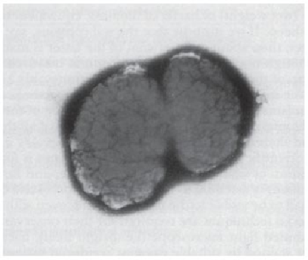
F
IG. 41
Two cells of a
Nitrosolobus
sp. in division; a nitrifying bacterium isolated from a Sri Lanka tea soil.
(Electron microscope photograph by N.W., magnification x 50,000.)
Objects, such as, bacteria, must be supported on a collodion-coated fine metal
grid (usually copper) which is inserted into the microscope. Air is pumped out to pro-
duce a high vacuum and the image can be seen on the fluorescent screen. To achieve
sufficient contrast in the outlines of a bacterium, the specimen is shadowed by sput-
tering with heavy metal (lead, chromium or gold) heated electrically in a high vacu-
um. Alternatively, the specimen may be negatively stained with phosphotungstate. To
see the internal structure of a bacterium, very thin sections of the organism must be
prepared. The material is embedded in a hard polymerized plastic and sections are cut
using an ultramicrotome equipped with a diamond knife or even a sliver of plate glass.
Heavy metals are used in stains because of their capacity for scattering electrons. The
electron microscope is a valuable aid in studying the fine structure of microorganisms
and viruses.
To examine microorganisms with the ordinary light microscope, a variety of
staining procedures is used, but these need not concern us here. The appearance and
morphology of different microorganisms and their various properties and character-
istics are important in their identification. Motile bacteria owe their ability to swim
about to flagella, which are hair-like appendages so fine that they can only be seen
under the microscope after special treatments and staining processes; flagella can also
be readily observed with the electron microscope. The number and arrangement of