Geoscience Reference
In-Depth Information
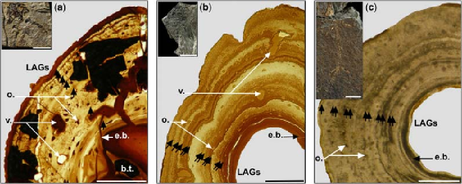
Fig. 2. Humeral diaphyseal bone histology of representative specimens among the largest of each Apateon taxa from
the Permian of the Saar-Nahe Basin, Germany. (a) Apateon pedestris (SMNS 54988, skull length SL 1.06 cm)
from Odernheim; (b) Apateon caducus (GPIM-N 1572, SL ¼ 1.6 cm) from Erdesbach and (c) Apateon pedestris (SMNS
55018, SL ¼ 0.76 cm) from Niederkirchen (b.t.: bone trabecula; e.b.: endosteal bone; LAGs: lines of arrested growth;
o.: osteocytes; v.: vascularization). Scale bars of thin sections: 0.1 mm; scale bars of fossil specimens: 1 cm.
remains free of trabeculae. The periosteal cortex
comprises primary bone tissue and the matrix is
essentially of lamellar bone. It contains numerous
round osteocytic lacunae, which are of the same
shape and size all along the cortex. The vasculariza-
tion, predominantly longitudinal (sometimes
radially arranged), is relatively dense and the
larger canals are mostly located in the deepest part
of the cortex. In the periphery of the marrow
cavity, a secondary endosteal deposition made of
lamellar bone is highlighted by a line of resorption.
The Katschenko's line is preserved between this
endosteal layer and the periosteal cortex.
Femur: Only femoral cross-sections of one
juvenile were available for this histological study.
The diaphyseal diameter is of 415 mm (GPIM-N
1680, Table 1). The cortex is not totally preserved,
preventing the complete cortical thickness, the
shape of the medullary cavity and the amount of
remodelling from being estimated. Even if only
the peripheral region of the cortex is still preserved,
it is nevertheless informative as it is constituted of
lamellar bone and remnants of calcified cartilage;
it shows relatively large and flattened osteocytic
lacunae but no vascularization.
only two in the humerus of SMNS 55018) run
longitudinally, essentially in the area situated at
mid-thickness of the cortex. The numerous oval
osteocytic lacunae are randomly localized. Against
the medullary wall, the endosteal coating is very
thin. There is no Katschenko's line.
Femur: The femoral diaphyseal sections are too
flattened to allow a significant estimation of the
diameter. Nevertheless, the cortex is relatively
thick (at 155 mm thickness in the largest individual
SMNS 55015), even if not completely preserved.
The innermost cortical region is not preserved in
any thin section. The preserved compacta is made
of lamellar bone and crossed by one longitudinal
primary vascular canal in SMNS 55015. The osteo-
cytic lacunae are flattened and homogeneously dis-
tributed in the periosteal cortex.
Differential diaphyseal bone-growth
features
The organization of different bone microstructures
is representative of the bone-growth rate such as
the density, size and shape of the cellular bone
lacunae and the density of vascularization (Amprino
1947). When the diaphyseal bone deposition is rela-
tively slow, bone cells are mostly flattened and the
vascularization is reduced or absent. When the dia-
physeal bone deposition is relatively fast, bone cells
are rounder, more numerous and the vascularization
is fairly denser. According to these criteria, it is
suggested that the mid-shaft bone deposition in the
stylopod long-bones of A. caducus was actually
faster than in the long-bones of both sets of A. pedes-
tris. The cellular shape would suggest a slightly
A. pedestris from Niederkirchen. Humerus (Fig. 2c):
Mid-shaft sections show diameters ranging from
500 to 580 mm (Table 1). The stylopod cortex is
rather thick at the diaphyseal level (up to 210 mm
thick in the largest individual SMNS 55018,
Table 1). The marrow cavity is therefore relatively
small and free from any medullary trabeculae. The
periosteal cortex, made of lamellar bone, is very
compact. Very few small primary osteons (e.g.