Biology Reference
In-Depth Information
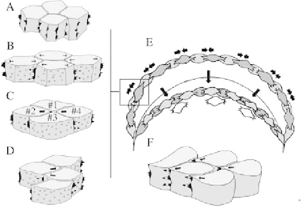
Figure 18.3 Mediolateral cell intercalation occurs as a result of polarized protrusive
activity. Before cell intercalation begins, deep mesodermal cells are pleiomorphic and their
lamelliform and filo-lamelliform protrusive activity (black protrusions in A-D) is
unpolarized in the plane of the tissue (A). At the onset of cell intercalation, these
lamelliform or filo-lamelliform protrusions become concentrated at the medial and lateral
ends of the cells and are thought to exert traction on adjacent cells (pointers, B-D). The
traction developed by these protrusions is thought to elongate the cells mediolaterally
(small arrows, B) and pull them between one another along the mediolateral axis (arrows,
C-D). These behaviours effectively form tensile arcs spanning the mediolateral aspect of
the prospective mesoderm, which are anchored at both ends near the vegetal endoderm (E).
It is the shortening of these arcs by cell intercalation (apposed arrows, E) that squeezes the
blastopore shut and aids involution (arrows, E). Deep neural cells are thought to
intercalate by using the same type of traction on one another, but in this case they appear
to do so by heavily biasing their lamelliform protrusive activity toward the midline (F). The
polarized cells are connected to one another at their anterior and posterior surfaces by
numerous, small contacts (dark grey patches, B-D)
or posteriorly. There they begin to exert traction on similarly displaced,
neighbouring cells located medial or lateral to them, and essentially
participate in a new arc (unshaded cells, Figure 18.3E). An important element
of this mechanism is that the cell body acts as a stable substrate on which the
lamelliform medial and lateral protrusions can exert traction (Keller et al.,
2000).
Neural cells also become polarized during mediolateral intercalation with
one major difference. Instead of being bipolar, with the lamelliform protrusive
activity orientated mediolaterally, this protrusive activity is heavily biased
toward the midline of the embryo (Elul and Keller, 2000) (Figure 18.3F). This
medially-directed protrusive activity is thought to drive cell intercalation by a
mutual cell-on-cell traction, similar to that seen in the bipolar mode (Keller