Biology Reference
In-Depth Information
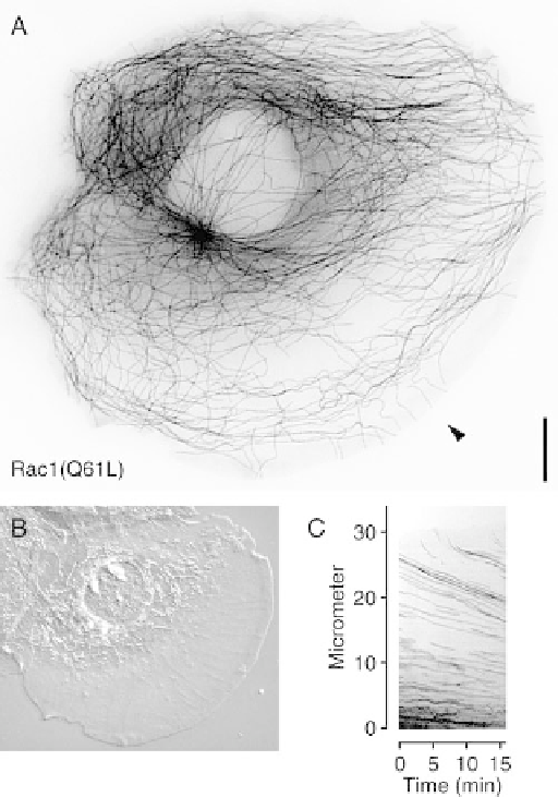
Figure 13.4 Microtubules in a Rac1(Q61L)-expressing PtK1 cell. (A) Microtubules in a
live PtK1 cell expressing dominant active Rac1(Q61L) observed by fluorescence
microscopy after injection of fluorescently labelled tubulin. The image was contrast
inverted and subjected to an unsharp mask filter to better visualize individual microtubules.
Dominant active Rac1(Q61L) induces lamellipodia formation all around the free cell edge
and many microtubules grow to the very edge of the cell although they are rapidly carried
away from the edge by actin-based retrograde flow. Bar, 10 mm. (B) Differential
interference contrast image of the same cell. (C) Kymograph analysis (time versus
distance plot) of the microtubule time-lapse series along the line indicated by the arrowhead
in (A). Oblique dark lines demonstrate microtubule retrograde flow
Rac1 and Cdc42 leads to Pak1-mediated phosphorylation of Op18/stathmin
(Daub et al., 2001) (Figure 13.3d). Indeed, phosphorylation of Op18/stathmin
upon stimulation with phorbol ester, another stimulator of Rac1 activity, had
been observed earlier (Hailat et al., 1990). Op18/stathmin is a tubulin-binding
protein that sequesters tubulin dimers and promotes microtubule plus end
catastrophes. Phosphorylation inactivates Op18/stathmin, which would