Image Processing Reference
In-Depth Information
4 Modifying a Resected Region Considering Hepatic
Veins
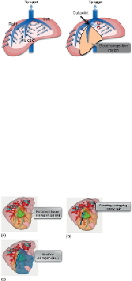
For preoperative planning, not only portal veins but also hepatic veins should be considered
due to a blood congestion problem by cuting hepatic veins.
Figure 7
explains the blood con-
gestion problem. There are three large hepatic veins in the liver: right, middle, and left hepatic
veins, and their major function is to carry blood from the tissue back to the heart. Similar to
the tumor metastasizing model described in
Section 1
, each hepatic vein is thought to absorb
blood from its closest tissue as shown in
Figure 7
. If a hepatic vein is cut at a point as shown
in
Figure 7(b)
,
blood congestion occurs in the region related to the end part from the cut-point
since the end part cannot absorb the blood; this blood-congestion region is damaged, and its
liver function reduces significantly. Hence, the congestion region is thought to be equivalently
a loss of the liver as well as a resected region. Therefore, if the total volume of the congestion
region and the resected region is large for the liver function of the patient, it would cause liver
failure. If the hepatic veins overlap with a portal-vein-based resected region, the resected re-
gion is limited so as not to overlap with any major hepatic vein.
FIGURE 7
Blood congestion problem.
In conventional software, surgeons must draw complex cuting surfaces manually consider-
ing hepatic veins, and it is very time-consuming since 3D surface is difficult to draw and not
accurate.
To solve this problem, we extend the portal-vein-based estimation so as to consider hepatic
estimation (
Figure 8(a)
). The yellow region denotes the portal-based cut region. Next, it is
checked whether the resected region overlap with hepatic veins or not. If overlapping (
Figure
8(b)
)
, the original resected region is limited so as not to overlap with the hepatic vein (
Figure
8(c)
). This modified resected region is denoted by the blue region.
FIGURE 8
Modifying the portal-vein-based resected region considering hepatic veins.
5 Conclusion
In order to minimize the resected volume while preventing metastases, the automatic method
that finds the optimal combination of cut-points is proposed. The domination ratio is a key to
success to find such optimal combination in a systematic way. Recently, the integration of 3D
simulation and ultrasonography is progressing to support the surgical operations in the real
Search WWH ::

Custom Search