Image Processing Reference
In-Depth Information
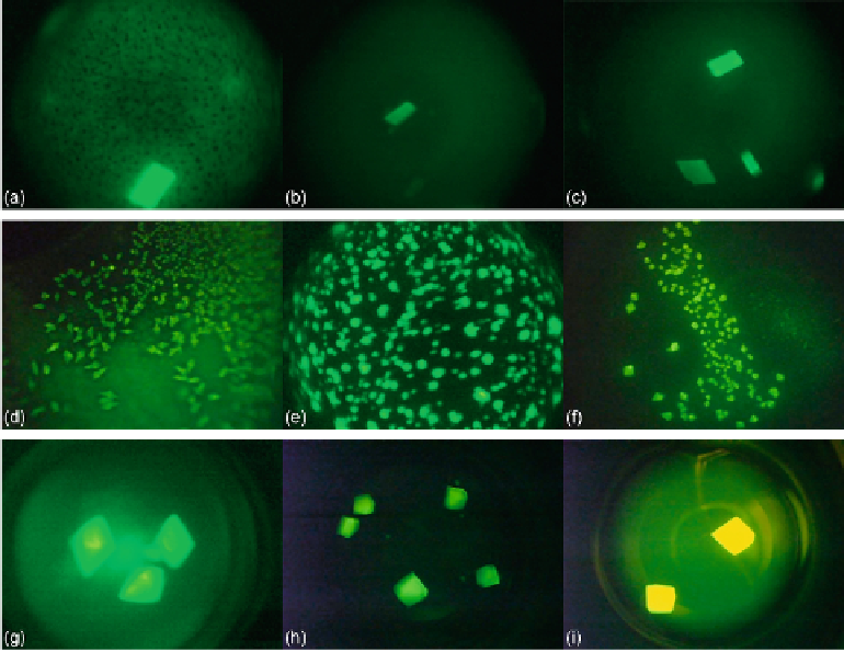
FIGURE 5
Protein crystallization image samples: (a-c) 2D plates, (d-f) small 3D crystals, and
(g-i) large 3D crystals.
4.1.2 Small 3D crystals
The areas of small 3D crystals are smaller than those of large 3D crystals. They have higher
intensities than 2D plates. This causes a significant intensity change between 3D objects and
background in images. Generally, it is hard to detect all the edges of this category due to small
size.
Figure 5
(d-f) shows some sample images of this category.
4.1.3 Large 3D crystals
This category generally has regions with high intensity, and these regions generally have
proper geometric shapes. The 3D structure of large 3D crystals can be observed in images. In
some particular cases, it is difficult to detect all the edges of 3D objects because of focusing
and light reflection problems. The instances of this category have larger sizes than small 3D
crystals. Some sample images of this category are shown in
Figure 5
(g-i).
4.2 Correctness Measurement
Most image binarization studies need to deal with a common difficulty regarding the cor-
rectness of their proposed system. Because a simple visual comparison of the binary images
would not provide an objective and dependable results, numerical results are expected as well
as the visual results. For this reason, we decided to generate a reference (ground-truth) bin-
ary image of each protein image manually in our dataset. We extracted the protein instances
Search WWH ::

Custom Search