Image Processing Reference
In-Depth Information
The process of rebuilding starts with applying a band-pass filter to the RF signal to eliminate
signals that do not come from the transducer. In the next step, a time gain compensation (TGC)
function is applied to compensate for atenuation loss. After this, the envelope of the signal is
computed and the result is log-compressed and normalized in a grayscale image.
After the process of rebuilding, the grayscale image, in polar coordinators, is submited to a
digital development process (DDP) responsible for enhancing the contrast and edge emphas-
is. So, the image is interpolated to cartesian coordinators. The cartesian image is further pro-
cessed with an intensity transformation function to improve the contrast of the final cartesian
grayscale IVUS image.
The above processes are described in more detail in
Section 2
and the results obtained are
DICOM images from an examination.
Section 4
also presents the conclusions and possibilities
for future work. This article is an extended version of the paper presented in the 2014 Interna-
2 Method for IVUS image reconstruction
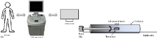
An IVUS examination is carried out by inserting a catheter into coronary arteries via femoral
or brachial vessels. At the tip of this catheter there is an ultrasound emiter and a piezoelectric
transducer that collect the echoes reflected by internal structures of the vessel as RF signal.
A schematic representation of the execution of an IVUS examination is shown in
Figure 1(a)
,
where the IVUS equipment collects data from patient and stores it in the workstation.
Figure
1(b)
shows an IVUS rotational catheter.
FIGURE 1
(a) IVUS
in vivo
analysis typical scenario and (b) rotational IVUS catheter.
During an IVUS exam, the acquired images are stored in DICOM format and exported to
the databank of the clinical centre to be used for clinical diagnosis.
To improve the quality of the images, in terms of CNR, physicians frequently need to adjust
parameters like contrast and brightness to improve the visualization of the region of interest
(ROI).
In addition to the images in DICOM format, the equipment allows the RF signal to be recor-
ded, which are used in the manufacture of images in a proprietary format.
The proposal of this article is to process the RF signal data according to the steps shown in
Figure 2
and detailed below, to rebuild the IVUS images with an independent and fixed set of
parameters.
Search WWH ::

Custom Search