Biology Reference
In-Depth Information
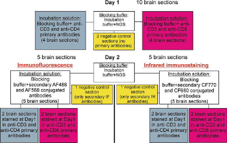
Fig. 4. Flow chart for performing double immunohistochemistry. The fl ow chart refl ects the methodological timeline and
steps to perform double-labeling immunohistochemistry.
See
text for detailed step-by-step explanations.
11. Incubate staining and negative control sections overnight at
4°C on shaker.
Note
: Antigen retrieval is not required for free-fl oating sections
because the brain tissue was not exposed to high formalin concen-
trations. In our example, as the CD protein concentration is low,
we used a 0.3% Triton-X 100 in PBS during overnight incubation.
The Triton-X 100 concentration and length of exposure is subject
to change; it is believed, the high Triton-X 100 concentration or
overexposure may increase background noise. The normally used
concentration range is 0.1-0.3%; the incubation time varies.
II. Day 2
(Table
4
)
Protocol
1. Using a paint brash, transfer sections into 24-well plates loaded
with 1 ml of Wash buffer in each well.
2. Rinse sections three times, 10 min each in Wash buffer on 3D
rotator.
3. To prepare blocking buffer (see calculation samples in
Table
4
):
(a) Take Incubation buffer (0.3% Triton-X 100 in Wash
buffer).
Search WWH ::

Custom Search