Biology Reference
In-Depth Information
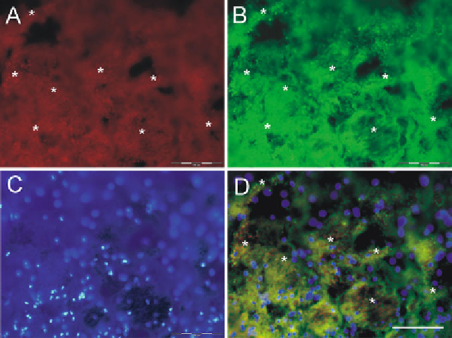
Fig. 3. Fluorescent microscopy of “naïve” brain sections. Excitation with a visual light spectrum may result in emission of
autofl uorescent signals which are normally stronger in aged or injured tissue. At day 1 post-ICH extravasated erythrocytes
are clearly visible in both red (excitation 530-550 nm,
a
) and green (excitation 470-490 nm,
b
) channels in “naïve” — no
staining with primary and or secondary antibodies. Cover slips with DAPI identifi ed intact cell nuclei (blue channel, excita-
tion 360-370 nm,
c
). As enucleated, erythrocytes are DAPI negative (
d
)
—
merge
a
,
b,
and
c
images. Asterisks (
a
,
b
and
d
)
indicate clusters of erythrocytes.
Bar
100
μ
m .
3. Pipettes.
4. Pipette tips.
5. Sharpie marker.
6. Wash buffer: 0.05% Tween in 0.1 M PBS, pH 7.4 (see recipe
below).
7. Incubation buffer: 0.3% Triton-X 100 in Wash buffer (see recipe
below).
8. Syringe fi lters Millex-FG, 0.20
μ
m cat. #SLFGR04NL.
9. Black tray.
10. Glass slides. No special brand preferences, but slides should be
compatible for use in fl uorescent microscopy (for example,
VWR Micro slides Superfrost Plus, VWR cat. No 48311-703).
11. Blocking serum (use the normal serum from the same host
species as the labeled secondary antibodies; in our example it is
a normal goat serum, NGS).
12. Primary antibodies.
Search WWH ::

Custom Search