Biology Reference
In-Depth Information
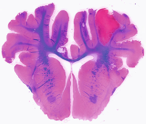
Fig. 4. Luxol fast blue staining showing white matter injury 3 days after ICH in pigs.
Measure the bilateral blue area.
Measure three times and get a mean value.
3.5.4. Pig White Matter in
Luxol Fast Blue-Stained
Brain Coronal Sections
(Fig.
4
)
●
●
Express the result as a percentage of the ipsilateral/
●
contralateral.
4. Notes
4.1. Animal Perfusion
and Sections
During the process of animal intracardiac perfusion, make sure
no air bubbles in the perfusing tubes.
When coverslip with Permount, leave no bubbles and keep the
●
●
slides clear.
4.2. Luxol Fast Blue
Staining
When differentiating the slides in the lithium carbonate solution
and continuing in the 70% ethanol, one can check under micro-
scope to see if gray matter is clear and white matter is sharply
defi ned. If not, repeat the differentiation steps in lithium carbon-
ate, 70% ethanol and rinse in distilled water. Neuron will change
from pink to violet, while myelin, including phospholipids from
blue to green.
Search WWH ::

Custom Search