Biology Reference
In-Depth Information
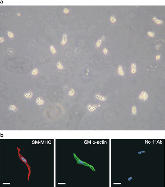
Fig. 3. Freshly isolated native cerebral artery myocytes. (
a
) Cerebral arteries were
enzymatically dissociated to obtain isolated vascular smooth muscle cells. Image shows
individual smooth muscle cells using differential interference contrast microscopy (x40).
(
b
) Vascular smooth muscle can be identifi ed by spindle-shaped cell morphology as well
as immunostaining of smooth muscle alpha actin and smooth muscle myosin heavy chain.
Scale bars
represent 10
μ
m. From Nystoriak et al. (
28
) with permission.
80
L
-glutamic acid (mono-sodium salt), 2.0 MgCl
2
, 2 CaCl
2
, 10
HEPES, and 10 glucose (pH 7.3; 37°C; 30 min) using a 1 ml vial
placed in a water bath. Arteries are then transferred to a Ca
2+
-free
GIS containing papein (0.3 mg/ml) and 1,4-dithioerythriol
(0.7 mg/ml; 37°C; 17 min) using a fi re-polished Pasteur pipette.
Next, arteries are incubated in GIS containing collagenase type F
(0.7 mg/ml), collagenase type H (0.3 mg/ml), and 100
M CaCl
2
(37°C; 20 min). Finally, arteries are incubated in GIS containing
2 mM CaCl
2
on ice (3×; 10 min) and gently triturated into indi-
vidual cerebral artery myocytes using a small bore fi re-polished
Pasteur pipette. Cerebral artery myocytes can easily be identifi ed
by characteristic spindle-shaped morphology (Fig.
3
). The cells are
kept on ice and used within 6 h of isolation.
μ
Search WWH ::

Custom Search