Biology Reference
In-Depth Information
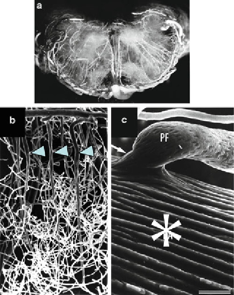
Fig. 3. (
a
) Macroscopic view of cast of the canine pons. (
longitudinal view
). (
b
) Scanning
electron microscopy at canine cerebral cortex. (
Arrow heads
: intraparenchymal arterioles).
(
c
) Not only microvessels but also major artery such as the basilar artery (*) can be
observed in detail.
kept in a freezer for several hours in order to promote
solidifi cation of a solution with gelatin. After that, the
brain is removed and kept in the fi xative solution used as
perfusion fi xative solution for 1 or 2 weeks. The brain is
sliced on the thickness of 1 mm or 2 mm using a microslicer,
and this slice is immersed in methyl salicylate for several
days or glycerol for 24 h until the brain tissue becomes
transparent. And this section is observed under a substance
microscope or a light microscope.
(c) Microangiography (Fig.
4b
, c) (
7
)
A solution of 30-50% of microbarium (micropaque,
baroperse, chromopaque, etc.), 5-20% of gelatin in distilled
water is used as a contrast medium of the blood vessels.
The solution is injected through the same cannula used in
Search WWH ::

Custom Search