Biology Reference
In-Depth Information
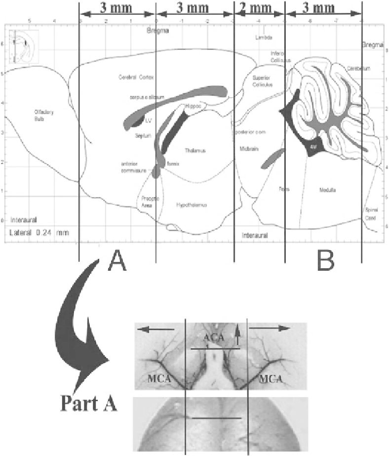
Fig. 1. Schematic representation of tissue cutting and embedding for the whole brain.
Section A
is for detecting the histo-
logical changes of middle cerebral artery (MCA) and anterior cerebral artery (ACA).
Section B
is for measuring basilar artery
(BA) cross-sectional area. The
lower panel
shows the ventral and dorsal images of
Section A
of the
upper panel
. Three cuts
were made in this section of the brain for obtaining perpendicular sections of the MCA and ACA, respectively. The
arrows
indicate slice direction.
3.4. Light Microscopy
or Slide Scanning
Place the slide on the microscope stage, with the coverslip facing
the source of light.
Adjust magnifi cation to 20-25 times (taking into account internal
camera magnifi cation).
Locate the artery of interest (BA, MCA, or ACA).
Search WWH ::

Custom Search