Biology Reference
In-Depth Information
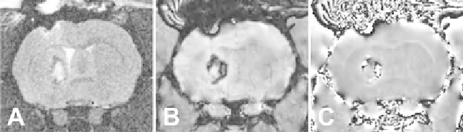
Fig. 3. Rat intracerebral hemorrhage shown on (
a
) T2 (
b
) SW image and (
c
) SWI phase. The SWI is more sensitive to the
blood products in the lesion while the T2 shows the edema triggered by the bleed.
images, can delineate the sequence of events as extravascular blood
is degraded within the brain (
34
). Since SWI is a new technique
that requires additional fi ltering and postprocessing that are largely
not yet available commercially from imaging manufacturers, we
delineate a detailed protocol for acquisition, processing, and analy-
sis of SWI-derived SAH data. Unequivocal imaging modalities,
such as SWI will greatly spur both clinical diagnosis and advances in
animal research allowing effective evaluation of potential therapies.
2. Materials
The protocol outlined below can be used for any animal model of
SAH, irrespective of the induction method. Additional instru-
ments, materials, and research apparatus may be required for the
induction of SAH. See Note 1.
2.1. Induction
of Anesthesia for
Neuroimaging
1. Oxygen gas tanks.
2. Isofl uorane vaporizer and induction box.
3. Isofl uorane.
4. Instrument with which to perform the pedal refl ex test (appli-
cation of pressure on a rat's hind limb ankle joint results in the
foot being withdrawn; refl ex is absent in deeply anesthetized
animals).
2.2. Imaging
Equipment
To confi rm the presence of an SAH lesion CT and/or MRI modalities
can be employed. In addition, these modalities may pose a research
benefi t to the study depending on the focus of the project by the
addition of quantitative imaging data. However, since we believe that
SWI imaging is superior to CT and other conventional MR modalities
(i.e., T2WI), we focus on acquisition of SWI. See Note 2.
When performing MRI on mice, one can use a Bruker Advance
11.7 T MRI (8.9-cm bore) with a 3.0 cm (internal diameter)
volume radiofrequency coil (Bruker Biospin, Billerica, MA) or
Search WWH ::

Custom Search