Biology Reference
In-Depth Information
multicontrast magnetic resonance imaging (MRI) sequences have
allowed noninvasive determination of the progression of this dev-
astating disease. While the purpose of this chapter does not provide
a comprehensive overview of neuroimaging of SAH, there are
reviews that adequately describe the current state of the art for
clinical management of the SAH patient (
1
). Provided in more
detail below are brief overviews of the primary imaging modalities
for SAH, a how-to for an MR-based acquisition and more
importantly analysis of MR datasets for rodent models of SAH.
The assessment of vasospasm by CT or MR angiography is not
discussed (see refs. (
3, 4
) for details).
1.1. Computed
Tomography
Computed tomography is often the fi rst imaging modality used to
clinically diagnose SAH. There are a number of reasons for this,
including: (1) wide-spread availability, (2) ease of use, (3) rapid
acquisition of data, and (4) early detection of blood products
(Figs.
1
and
2
). CT is a highly accurate test; however, it suffers
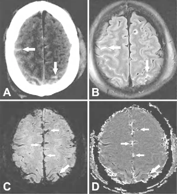
Fig. 1. (
a
) CT
arrows
indicate SAH. (
b
) Corresponding FLAIR image with
arrows
indicating
SAH. SAH is shown similarly on FLAIR and CT. (
c
) SW image (
d
) SWI phase image. The SWI
images highlight SAH (
arrows
) in different regions than the FLAIR adding complementary
information. Images courtesy of Zhen Wu.
Search WWH ::

Custom Search