Biology Reference
In-Depth Information
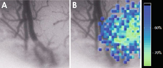
Fig. 5. LASCA image of focal ischemia. (
a
) The
upper image
gives a conventional surface picture. Note the virtual
disappearance of pial arterioles on the right side of the picture due to focal vasospasm in the microcirculation. (
b
) The
pseudo-colored image
shows the rCBF reduction in the cortex on the right side.
laser light with slightly different path lengths. A granular or speckle
pattern is generated when an area illuminated by laser light is
imaged onto a camera. A time-varying speckle pattern is produced
at each pixel in the image if the scattering particles are in motion.
The variations of this pattern contain information about the motion
of the scattering particles and quantitative (relative) fl ow informa-
tion is obtained through measuring the temporal and spatial inten-
sity fl uctuations of the speckles and conversion of the raw speckle
images by specifi c analysis algorithms into two-dimensional blood
fl ow maps (
33, 37
). As such, LASCA is not based on a spectral
analysis of scattered laser light like LDF but images and evaluates
the spatial changes of speckle fl uctuations. It has been used to eval-
uate rCBF changes in response to functional activation, SD, and
ischemia in cranial window preparations or through the thinned
skull (
34, 46-49
). An important advantage over LDF is the ability
for two-dimensional monitoring of rCBF changes (Fig.
5
). The
possibility to combine speckle imaging with fl uorescence imaging
or optical spectroscopy (
50, 51
) offers the option for multiparam-
eter brain imaging. Like LDF, LASCA does not allow quantifi ca-
tion of rCBF in absolute units. Another limitation comes from the
fact that the signal is dominated by superfi cial fl ow changes and
gives limited information on changes in deeper cortical layers.
4.3. Optical Imaging
of Intrinsic Signals
and Optical
Spectroscopy
Optical imaging methods are frequently used for studies of neuro-
vascular coupling. The general assessment of refl ectance changes as
a wave of SD passes to a brain slice is described in Chap.
29
.
This
allows easy identifi cation of localization, spread, and speed of SD
waves. OIS is also widely used in the blood-perfused brain and the
general principle is based on the correlation of changes in light
refl ectance with changes in neuronal activity due to changes in
Search WWH ::

Custom Search