Biology Reference
In-Depth Information
5.2. Two-Photon Laser
Scanning Microscopy
Modern imaging techniques, such as two-photon laser scanning
microscopy (2PLSM) (
45
), have greatly contributed to our under-
standing of cellular physiology and dynamics of many cell types in
brain slices in vitro as well as in in vivo preparations. Imaging with
2PLSM is also useful for high-resolution cellular monitoring dur-
ing SD. With 2PLSM, problems associated with scattering and
absorption of photons by neural tissue are signifi cantly reduced as
compared with one-photon excitation used in confocal and wide-
fi eld fl uorescence microscopy (
46
). In 2PLSM, the use of the long-
wavelength excitation photons results in less scattering and low
absorption by water and intrinsic tissue chromophores and pro-
vides better tissue penetration. Simultaneous absorption of two
photons that combine their energies excites a chromophore and
generates fl uorescence that can be acquired from several hundred
microns deep within living tissue. The intensity-squared require-
ment of two-photon excitation restricts chromophore excitation
and emission to a narrow focal plane (focal slice), where the laser
beam is focused. This reduces photobleaching and photodamage
in thick living tissue, permitting repeated sampling (
47-49
). The
intrinsic confocality of two-photon excitation results in thin optical
sections that can be stacked to create a 3D image.
In recent years, generation and availability of transgenic mice
expressing fl uorescent proteins in a small percentage of brain cells
have greatly facilitated imaging by providing the opportunity to
capture detailed time-lapse images to observe the dynamic proper-
ties of living cells. Here, we summarize how we have used 2PLSM
in live cerebral slices from transgenic mice expressing the green
fl uorescent protein (GFP) to visualize single-fl uorescent neurons
and astrocytes responding to SD in real time, at high magnifi cation
and deep in intact tissue (Fig.
3
) (
50
). We have made cerebral slices
from transgenic mice expressing GFP in a small subset of pyramidal
neurons in cerebral cortex and hippocampus (
51
) and from mice
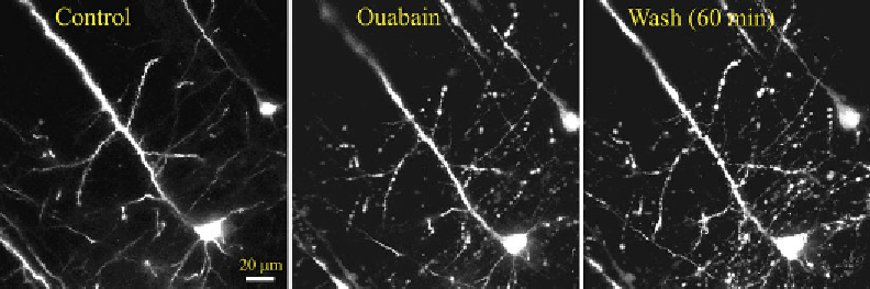
Fig. 3. No recovery from dendritic beading with spine loss and neuronal somata swelling after 1 h of washing out in normal
aCSF following SD triggered by 100
μ
M ouabain application. Images are MIPs of several optical sections acquired <100
μ
m
below the slice surface.
Search WWH ::

Custom Search