Biology Reference
In-Depth Information
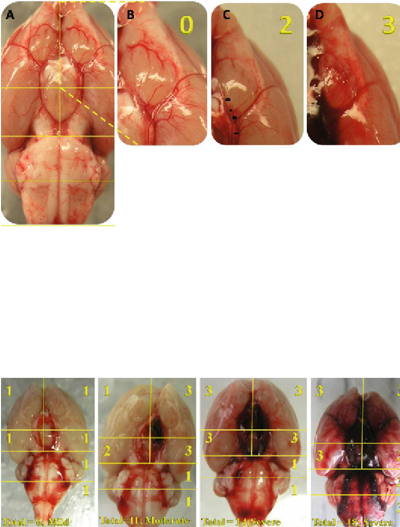
Fig. 2. Example of the grading system using examples of the left frontal basilar cistern as an example. (
a
) Shows the brain
from an SHAM animal. (
b
) Close up view of the left frontal basilar cistern shows clearly visible vessels without subarach-
noid blood. The grade for this section is 0. (
c
) The left frontal basilar cistern from an SAH animal. This time the cistern has
a signifi cant blood clot but it does not obscure the ICA and ACA arteries. The grade for this cistern is 2. (
d
) Left frontal basilar
cistern of another SAH animal. This time there is severe SAH, obliterating the view of all vessels. The grade here is 3. Photo
adopted with modifi cation from Sugawara et al. (
11
) .
Fig. 3. Here are four examples of various SAH grades ranging from mild to severe created using the endovascular perforation
model in rats. The conditions for each animal were under identical and under controlled conditions. These photos represent
the variability inherent in the endovascular perforation model.
Search WWH ::

Custom Search