Biology Reference
In-Depth Information
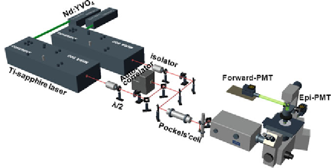
Fig. 2. Schematic of a CARS imaging setup.
1,000 nm, where water absorption is minimized. The pulse
width is 2.5 ps whose corresponding spectral width matches
the line width of Raman bands. The laser source for CARS can
also be realized by using optical parametric oscillator (OPO)
pumped by a pulsed laser (
16
).
2. Optical pathway: In Fig.
2
, collinear beam geometry is utilized
to fulfi ll the phase matching condition which also simplifi es
the implementation. The two laser beams are tightly synchro-
nized (Sync-Lock, Coherent Inc) with an average timing jitter of
100 fs. The two beams are collinearly combined using a dichroic
combiner (LWP-45-R720-7850-PW-1004-UV, CVI Laser
LLC, Albuquerque, NM). A Pockels cell (Model 350-160,
Conoptics, Danbury, CT) is used to lower the repetition rate,
which reduces average power but maintains high peak power
at the sample. The pockels cell is important in the imaging
of fi xed slices where a low laser power is needed to minimize
the photodamage. In the imaging of fresh tissues and live
animals where higher laser power is required, the pockels cell can
be removed from the optical pathway. The polarization of the
incident beams is controlled by half-wave plates.
3. Laser-scanning microscope: Two collinearly combined beams
are directed into a laser scanning confocal microscope (FV300/
IX70, Olympus America Inc, Melville, New York). To produce
a high-quality CARS image, a high numerical aperture (NA)
objective, such as a 60× water objective (NA = 1.2) or a 40× water
objective (NA = 0.8) is used to form tightly focused excitation
beams through the sample. To obtain a CARS image in a large
fi eld of view, a 20× air objective (NA = 0.75) is also acceptable
to generate CARS signal.
Search WWH ::

Custom Search