Biology Reference
In-Depth Information
exogenous T1 contrast agents such as gadolinium-DTPA do not
cross the intact blood-brain barrier (BBB), systemic injection of
such agents can be used to evaluate BBB disruption in TBI models
(
5
). Another widely used imaging sequence, T2-weighted imag-
ing, is particularly useful for assessing edema and encephalomalacia
after TBI (
1, 6
). The altered physical environment of water in
edematous tissue makes these areas appear hyperintense (brighter)
compared to surrounding tissue. In contrast, hemorrhage and
contusions are typically visible as regions of hypointensity on
T2-weighted imaging. A third approach, T2*-weighted imaging,
is also highly sensitive for visualizing hemorrhage (
4
). In practice,
a combination of MR images using different image weighting will
likely yield more complete information than a single imaging
sequence (Fig.
1
).
Both qualitative and quantitative evaluations of TBI pathology
can be carried out using MR imaging. By measuring the area of a
traumatic lesion on a series of MR images and multiplying by the
image slice thickness, a good estimate can be made of the lesion's
total 3-dimensional volume. Lesion volumes calculated with this
technique strongly correlate with histological estimates of lesion
volume (
1, 7, 8
). However, using MRI to assess TBI lesions offers
distinct advantages over histological methods. Fewer animals are
required, since there is no need to sacrifi ce separate groups at each
time point. Moreover, the potential variability inherent in a cross-
sectional study design is reduced. Investigators can track the effects
of TBI noninvasively as the lesion pathology develops. Perhaps
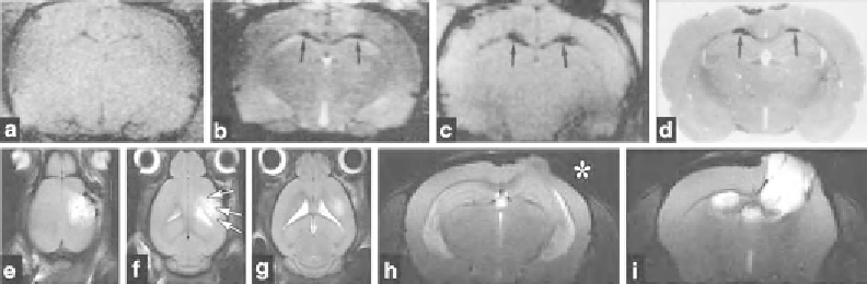
Fig. 1. MR image weighting options in animal models of TBI. (
a
-
c
) Coronal images of a rat brain taken at 4.7 T, 2 days after
midline fl uid percussion injury. (
a
) T1-weighted MRI (TR = 500 ms; TE = 15 ms); (
b
) T2-weighted MRI (TR = 3,000 ms;
TE = 80 ms); (
c
) T2*-weighted MRI (TR = 350 ms; TE = 10 ms; fl ip angle, 20°). (
d
) A corresponding H & E stained histological
section from the same brain. Arrows point to hemorrhage in the corpus callosum. (
e
-
g
) Horizontal T2-weighted images of
a mouse brain taken at 9.4 T, 2 days after controlled cortical impact injury. Arrows indicate tissue edema at the lesion site.
(
h
-
i
) Coronal T2-weighted images (TR = 2,500 ms; TE = 45 ms) of a mouse brain taken at 9.4 T, 28 days after controlled
cortical impact injury. The increased spatial resolution resulting from the higher magnetic fi eld is apparent (compare to
a
-
c
). At 28 days, the T2-weighted images reveal enlarged lateral ventricles and the fl uid-fi lled cavity that has developed
at the injury site. An asterisk indicates cortical thinning ipsilateral to injury. Images are reproduced from Iwamoto et al.
1997 with permission from Wolters Kluwer and from Onyszchuk et al 2008 (
28
) .
Search WWH ::

Custom Search