Biology Reference
In-Depth Information
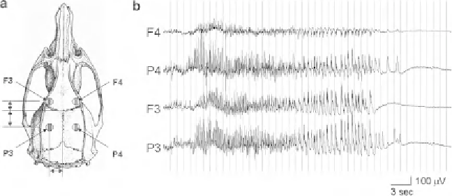
Fig. 3. Example of an EEG recording from the status epilepticus animal model. (
a
) A scheme of four recording electrodes
(F3, F4; P3, P4) was placed on the surface of skull. (
b
) The EEG recording shows a typical case of pilocarpine-induced
status epilepticus (Stage-I).
to allow rats to turn freely without twisting the cable. Following a
baseline EEG recording of at least 1 h (Fig.
3
), SE was induced by
injecting pilocarpine (30 mg/kg, i.p.). EEG recordings were used
to time the sacrifi ce depending upon the stage of SE desired.
Control rats were not given pilocarpine.
In this study, we ask whether selective C-fi ber input to cerebellar
cortex evokes a specifi c “slow-pain” response. The experimental
procedure was the same as our previous description (
12, 13
).
Briefl y, adult cats were anesthetized using
3.2.2. EP Responses
in Cat Cerebellar Cortex
Evoked by a Selective
Stimulation of Peripheral
C-Type Nerve Fiber
-chloralose (60 mg/
kg, i.v.). The trachea was canulated and both anterior and posterior
lobes of cerebellar cortex were exposed after partial removal of the
bony tentorium and occipital bone. The saphenous nerve was
dissected free and its arterial blood supply left intact while the
distal end was ligated and cut. Bipolar stimulation and recording
electrodes were placed on the centripetal portion of the nerve from
distal to proximal for monitoring of action potential volleys.
A Ag-AgCl blocking electrode was placed between stimulation
and recording electrodes to block A-fi ber inputs. During the course
of the experiment, the animal was paralyzed by injection with 4%
Flaxedil (20 mg/kg, i.v.) and artifi cially ventilated. The rectal tem-
perature and end-respiratory CO
2
were continuously monitored
and maintained within a normal physiological range. The saphen-
ous nerve was electrically stimulated 20 times with single pulses
(0.5 Hz and 0.2 ms) to obtain an averaged evoked potential. The
stimulus strength was adjusted as a multiple of the threshold inten-
sity (T) of A-type fi bers as determined from nerve volleys. The
intensities of the polarizing currents to selectively block A-fi ber
conduction were 30-120
α
A.
Figure
4
shows an example of how selective activation of
C-fi bers induces an evoked potential in the cat cerebellar cortex.
Stimulation of the saphenous nerve at 4 T strength, which excited
μ
Search WWH ::

Custom Search