Biology Reference
In-Depth Information
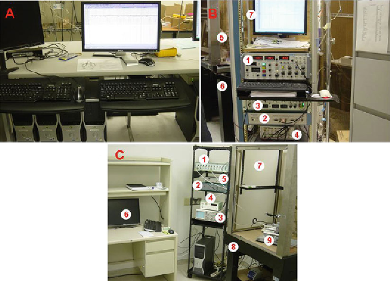
Fig. 1. Electrophysiological equipment for EEG, EP, and extracellular single-unit recordings. (
a
) Representative of a digital
EEG Comet XL Lab-based PSG system from GRASS Technologies. (
b
) Representative of an electrophysiological recording
system from AxoClamp 2B from Axon Instrument (Fig.
1b
-1) with HS-2A headstage (Gain: ×1 LU) for evoked potential
recordings. (
c
) Representative of an electrophysiological recording system for extracellular single-unit recordings.
waveform which permits us to differentiate between dopaminergic
and nondopaminergic neuron fi ring. Our oscilloscope is manufac-
tured by Hitachi (VC-6524, Fig.
1c
-3). In addition, an audio system
is used to monitor membrane potential changes caused by action
potential fi ring (Fig.
1c
-5).
2.1.3. Stimulator
We use A-M System isolated pulse stimulator (Model 2100) for EP
study (Fig.
1b
-3). It can be used in either voltage- or current-
stimulation modes.
A computer is used for both data acquisition during experiments
and data analysis thereafter (Fig.
1a
, b-7, c-6). An analog-digital
converter is used so that signals can be recognized by the computer.
It is also used in reverse to send command signals from the computer
to the amplifi er. We use Digidata 1322A (from Axon Instrument)
for EP recording (Fig.
1b
-4) and Sky-A4 Bioelectric signal processing
system (Fudan University; Shanghai, China) for single-unit recording
(Fig.
1c
-2).
2.1.4. Computer and A-D
Converter
Search WWH ::

Custom Search