Biology Reference
In-Depth Information
Cg
As
As
(A)
(D)
Ld
Cld
Se
Lu
Pd
Cc
Md
Oe
Avg
Vg
(B)
(E)
Mx
Md
Pk
Ep
G
F
Sd
Fs
Ep
F
(C)
(F)
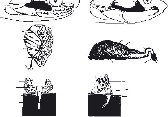
Figure 2.1
Comparison of a Duvernoy's gland system in an “opisthoglyphous”
(“rear-fanged”
non-front-fanged) snake (left) and a venom gland system in a model
“proteroglyphous” or “solenoglyphous” snake (right). Panel A. In the non-front-fanged
(“opisthoglyphous”) snake, Duvernoy's gland (shaded) is located in the temporal region.
Adjacent striated muscles (e.g.,
adductor superficialis
) run medially past the gland, but usually
are not directly attached.
Dispholidus typus
is an exception to this, as it does have limited muscle
attachment to the gland. Panel B. A cross-sectioned view of the Duvernoy's gland that reveals
the arrangement of the internal duct system draining the extensive parenchyma. A single duct
departs from a small, central cistern within the gland, and runs to a cuff of oral epithelium
surrounding the posterior maxillary tooth (F). Panel C. When the posterior maxillary tooth
penetrates the integument of the prey or human victim, the cuff of the oral epithelium remains
on the surface, thereby receiving Duvernoy's secretion, which flows around the tooth that may
(as depicted here) or may not be grooved (an “open system” with inherently low pressure).
Panel D. The venom gland (shaded) of this model proteroglyphous or solenoglyphous snake
includes a main venom gland, main duct accessory venom gland, and secondary duct that empty
into the base of the canaliculated (hollow) fang. Striated jaw muscles (e.g.,
adductor externus
superficialis
in “proteroglyphous” elapids or
compressor glandulae
in “solenoglyphous”
viperids) act directly upon the venom gland to raise the intraglandular pressure and send a pulse
of venom from the gland through the duct to the fang. Panel E. A sagittal view of the venom
gland reveals the secretory epithelium, and extensive storage reservoir of venom. Panel F. When
the fang penetrates the integument of the prey, the attachment of the venom duct to the fang
tightens in order to maintain the relatively high-pressure head, and venom passes down the
hollow core of the fang to be delivered deeply into the tissues (a “closed system” with inherently
high pressure). Abbreviations: jaw muscles,
adductor mandibulae externus superficialis
(As),
compressor glandulae
(Cg), accessory venom gland (Avg), central cistern (Cc), common lobular
duct (Cld), epithelium of prey integument (Ep), fang or enlarged maxillary tooth (F), fang sheath
(Fs), groove on surface of maxillary tooth (G), lobular duct (Ld), lumen holding secretory
product (Lu), main duct (Md), maxilla (Mx), oral epithelium (Oe), pocket of oral epithelium
around tooth (Pk), primary venom duct (Pd), secondary venom duct (Sd), secretory epithelium
(Se), main venom gland (Vg). After Weinstein and Kardong (1994) used with permission.