Biology Reference
In-Depth Information
(H)
(I)
(K)
(J)
Plate 4.20 (
Continued
)
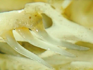
(H-K)
Dispholidus typus
, osteological specimen.
Note the multiple enlarged posterior
maxillary teeth with prominent lateral sharp ridges, and deep grooves running about 65%
of the length of the respective teeth. These adaptations, and a potentially wide buccal gape,
probably influence the lethal potential of bites by this species.
Plate 4.20E
, photo copyright to Dave Ball;
Plate 4.20
F and G, photos copyright to David
A. Warrell;
Plate 4.20
H-K, AMNH #75722, photos copyright to Scott A. Weinstein.
which took 5 weeks to heal (Knabe, 1939), and another had massive swelling with
blood-filled bullae (Spies et al., 1962). Local tender lymphadenopathy may develop.
Laboratory investigations confirm incoagulable blood, defibrinogenation, elevated
fibrin(ogen) degradation products, severe thrombocytopenia, anemia, and comple-
ment depletion via the alternative pathway (Nicolson et al., 1974; see also
Table 4.1
).
As there is no commercial antivenom for
Thelotornis
envenomations, a cas-
cade of symptoms often ensues. Patients may present with local edema, uncon-
trolled bleeding from the gums, and healing pre-existing wounds (Broadley, 1959;
Muguti and Dube, 1998), extensive ecchymoses, hematuria, coagulopathy, raised
fibrin(ogen)-related degradation products, thrombocytopenia, intravascular hemoly-
sis, multiple organ hemorrhage, and acute kidney injury [AKI; often manifested as
acute renal failure (ARF) characterized by acute tubular necrosis, probably primarily
from hemoglobinuric nephropathy; see Section 4.6] that is unresponsive to diuret-
ics or dialysis, such as occurred after Robert Mertens was bitten by a
T. kirtlandii
that resulted in death 18 days later (
Table 4.1
). Patients may also develop signs of