Biology Reference
In-Depth Information
morbidity from likely treatment-induced morbidity. This case emphasizes the essen-
tial need for adequate, objective, detailed documentation in cases of unusual and
severe envenoming, including formal medical evaluation by a physician, preferably
with experience in clinical toxinology (see Section 4.5). Publication of cases lack-
ing sufficient clinical information must be discouraged, particularly when they report
atypical serious effects of bites from species that are generally considered to have
mildly toxic Duvernoy's secretions, or are “mildly venomous.” Such anecdotal and
unvalidated case reports can create false perceptions of the potential hazards associ-
ated with bites from particular colubroid species (Section 4.5).
Although well-documented case reports suggest that bites from these snakes can
cause local medically significant bites resembling mild-to-moderate crotaline enven-
oming, as indicated previously, there are very few cases in which systemic enven-
oming could be inferred. An example of a patient bitten by a verified
P. olfersii
who developed systemic effects that were manifested by widespread ecchymoses is
shown in Plate 4.40A and B. Therefore, large specimens of
Philodryas
spp. should
not be carelessly handled in view of the known hemorrhagic, fibrinogenolytic, and
possibly neurotoxic properties of their Duvernoy's secretions. The medical signifi-
cance of bites from most species remains only partly characterized.
4.2.2.1 Overview of the Duvernoy's Gland and Associated Dentition of
Philodryas
spp.
The two
Philodryas
spp. that have received the most attention,
P. olfersii
and
P.
patagoniensis
, have enlarged posterior, grooved maxillary teeth. The posterior maxil-
lary teeth of
P. olfersii
have a deep groove occupying most of their length (Fry et al.,
2008).
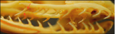
Philodryas baroni
has enlarged posterior maxillary teeth with deep grooves
present on almost the entire length of the teeth (Plate 4.38C-E). Although there is
limited information, it is likely that many, if not all, members of the genus exhibit
similar dentition.
There is also little information regarding the ultrastructure and functional morphol-
ogy of the Duvernoy's gland of this genus. However, Taub (1967) described the gland
(C)
Plate 4.38
(C and D) Skull and enlarged maxillary teeth of
Philodryas baroni
.
The teeth
are recurved, and the posterior maxillary teeth are significantly enlarged (indicated by arrows).
(E) Close-up of enlarged posterior maxillary teeth of
Philodryas baroni
.
There is a prominent
diastema between the anterior, and the notably enlarged, deeply grooved posterior maxillary
teeth. The grooves extend along almost the entire length of the teeth (indicated by arrows).
Plate 4.38A, photo copyright to Daniel E. Keyler; Plate 4.38B, photo copyright to Maik Dobiey;
Plate 4.38C-E, AMNH #62831, photos copyright to Arie Lev.