Biomedical Engineering Reference
In-Depth Information
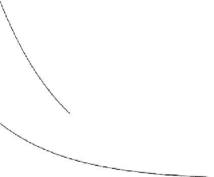
Iron Loading with Cell Proliferation
40
R
2
30
Ferucarbotran
M400
M500
M600
M750
0.992
0.990
0.999
0.995
0.993
20
10
0
0
2
4
Population Doubling
Fig. 2
Halving of cellular iron when MSC divide. The measurements of cellular iron with respect
to the number of times the cell count has doubled are shown as points. The R
2
values are the
exponential decay fittings to the points with a half-life of 1 population doubling. These results
show that when MSC are labelled with ferucarbotran or 400-750 nm MGIO, the iron mass per
cell approximately halves when the cells undergo one population doubling (unpublished data)
methods of identifying donor cells by means of FISH or sex mismatch may prove to
be an essential procedure to verify MRI observations in preliminary experiments.
7.3
Cellular Quantification
Quantification of cells through the use of radioactive methods such as scintigraphy
is well-established. Quantification of cells by iron oxide MRI on the other hand is in
its infancy, but reports to date have been promising. As particle diameter increases,
its relaxation rate increases and saturates at a constant value, as defined by the static
dephasing regime (SDR) (Lee et al.
2010b
). When cells compartmentalise small
particles such as SPIO or USPIO, their MR relaxation is similar to that of large
magnetic spheres operating in the SDR (Bowen et al.
2002
; Ziener et al.
2005
).
Since the relaxation rate of cells is constant when in the SDR, the R2* relaxation
rate is directly proportional to the number of cells per voxel when iron mass per cell
is known (Bowen et al.
2002
).
The accurate measurement of R2* relaxation rate is beset with artefacts from
macroscopic magnetic susceptibility of tissue-air interfaces, leading to overestimates


Search WWH ::

Custom Search