Biomedical Engineering Reference
In-Depth Information
Labelled stem cells, directly injected into myocardial infarcts can also be
monitored. Ferumoxide-labelled (Kraitchman et al.
2003
) and micron-size iron
oxide particles (MPIO)-labelled (Stuckey et al.
2006
; Hill et al.
2003
) allogeneic
MSC have been injected intramyocardially into infarcted hearts and their location
studied up to 16 weeks in various animal models. In a different study, intravenously-
injected iron oxide-labelled, allogeneic MSC homed to the myocardial infarct
in a canine model and persisted for at least 1 week (Kraitchman et al.
2005
).
However, the successful identification of hypo-intense cells may be complicated
by haemorrhage from microvascular obstruction during infarct reperfusion (van
den Bos et al.
2006
). Haemorrhage causes susceptibility-induced signal voids that
appear similar to iron oxide induced ones.
The homing of intravenously-injected MSC has been demonstrated in other
disease models as well. The arrival of ferumoxide-labelled MSC at injured arteries
(Gao et al.
2007
) and atherosclerotic plaques (Qiu et al.
2007
) was monitored by
MRI. The use of MRI tracking to optimise cellular delivery was also demonstrated.
Ferumoxide-labelling provided evidence of better MSC engraftment when cells
were given through the internal carotid artery, compared to intravenously in a
rodent stroke model (Walczak et al.
2008
).
A number of cell types have been shown to migrate towards the injured central
nervous system. MSC labelled with MPIO were shown to migrate towards white
matter (Chen et al.
2010
) or spinal cord injury (Gonzalez-Lara et al.
2010
).
Ferumoxide-labelled MSC (Jendelova et al.
2003
), ferumoxide-labelled neural
stem cells (NSC) (Zhu et al.
2006
) or USPIO-labelled ESC (Hoehn et al.
2002
)
were transplanted and shown to migrate from the contralateral hemisphere and
across the corpus callosum to the infarct periphery. By optimising MSC labelling
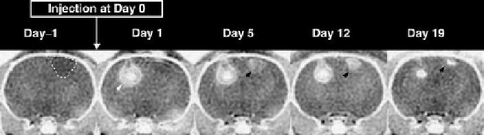
with 600 nm MGIO, the cells can be tracked in a rat stroke model using modest,
standard clinical hardware over 19 days (Fig.
1
) (Lee et al.
2009
). The same
Fig. 1
In vivo
MR tracking of MSC labelled with 600 nm MGIO to stroke lesion. A focal cortical
stroke (
dashed boundary
) was induced at Day 2 and 2 × 10
4
MSC were transplanted contralaterally
on Day 0 (
black arrowhead
). An area of hypo-intensity appeared in the stroke lesion (
white
arrowheads
) noticeable at Day 5, and increased over time to Day 19. Images were acquired with
gradient echo sequence - field of view: 5 cm; matrix: 160 × 160 and zero-filled to 512 × 512;
voxel dimensions: 313 mm × 313 mm × 1.5 mm; repetition time/echo time/flip angle/number of
excitations : 280 ms/20 ms/20°/15, as 10 two-dimensional slices (unpublished data)
Search WWH ::

Custom Search