Biomedical Engineering Reference
In-Depth Information
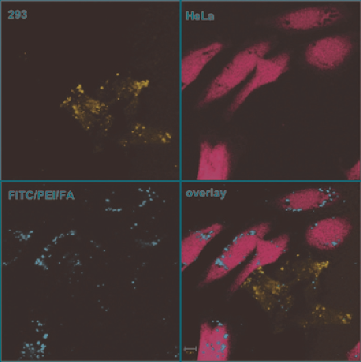
Fig. 10
Specific endocytosis of FITC/PEI/FolicAcid-functionalized silica nanoparticles in coculture
of HeLa and 293 cells. The cells were labeled with
blue
CMAC (HeLa) or CellTracker
Red
(293)
and plated together overnight prior to incubation with the particles for 4 h. After incubation the
extracellular fluorescence was quenched by trypan blue and the endocytosed particles with FITC-
label (
green
) inside blue- or red-labeled cells were detected by confocal microscopy. Scale bar
10 mm. (Reproduced with permission from (Rosenholm et al.
2009
). Copyright 2009 American
Chemical Society)
by enzymes in the lysosomes, the system can be exocytosed either directly or after
partial degradation, or the particles can accumulate inside the cells, potentially leading
to side effects such as formation of reactive oxygen species, affecting the cell cycle,
influencing cytokine synthesis… (Hu et al.
2009
). At this time, very few data has
been gathered on this topic. Fluorescent mesoporous nanoparticles have been shown
to escape endolytic vesicles and resist lysosomal degradation (Huang et al.
2005
).
In the case of biopolymer-silica hybrid nanoparticles, it was observed that gelatin or
alginic acid could be dissolved intracellularly within 3 T3-fibroblasts, leaving silica
nanograins in the vesicles (Boissière et al.
2006
; Allouche et al.
2006
). However,
nothing is known about further degradation/exocytose of these particles.
Search WWH ::

Custom Search