Biomedical Engineering Reference
In-Depth Information
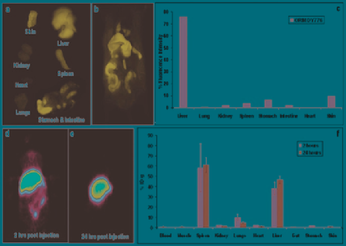
Fig. 3
Biodistribution of silica nanoparticles by the measurement of the fluorescence (
a
-
c
) and
radioactivity (
d
-
f
) from different organs of the mice injected with DY776 conjugated and
124
I
labeled silica nanoparticles respectively: (
a
) fluorescence acquired from the individual organs, (
b
)
whole body fluorescence imaging of the mice 24 h postinjection, (
c
) quantitative estimation of
fluorescence acquired from different organs of the mice, (
d
and
e
) PET images of the mice
injected with
124
I-silica nanoparticles 2 and 24 h postinjection, respectively, and (
f
) quantitative
radioactivity measurements from the individual organs of the mice. (Reproduced with permission
from Kumar et al.
2010
. Copyright 2010 American Chemical Society)
intraperitoneal infusion. On this basis, it was suggested that silica particles are 100
times more toxic than poly(lactic-co-glycolic acid) (PLGA) nanoparticles.
To close this section, it is worth noting that studies performed on zebrafish embryo
showed no uptake (silica particles being located on the chorion, a porous membrane
surrounding the fertilized egg) and therefore no mortality nor physiological deforma-
tion (Fent et al.
2010
). This enlightens an important issue related to possible silica
particle internalization through the skin, that is still to be elucidated (Park et al.
2010
).
3
Silica-Based Nanomaterials for Drug Delivery
3.1
Drug Encapsulation in Plain Particles
The simplest method is to entrap hydrophilic functional molecules within the silica
matrix
via
non-covalent interactions. This procedure is commonly used to incorporate
luminescent dyes into the silica matrix. The main drawback is the possible leakage of
Search WWH ::

Custom Search