Biomedical Engineering Reference
In-Depth Information
3.2.2
Composite Systems with Classical Polymersomes Charged
with Photosensitizers
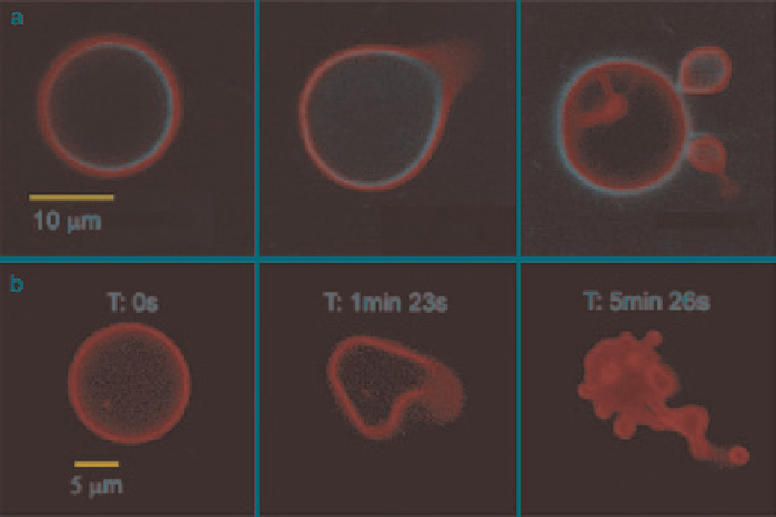
The group of Dmochowski has first reported photoinitiated destruction of composite
porphyrin-protein polymersomes (Robbins et al.
2009
). The polymersomes were
formed by incorporating a protein in the aqueous interior and a meso-to-meso ethyn-
bridged bis[(porphinato)zinc] (PZn
2
) chromophore in the membrane polymersomes
made from classical photo-inert PEO-
b
-PBD. Confocal laser scanning microscopy
(CLSM) imaging of polymersomes loaded with both ferritin and PZn2 at excitation
wavelengths (488, 543, or 633 nm, where PZn2 absorbs strongly) caused many of
the vesicles to undergo irreversible morphological changes ranging from formation
of new bends or “arms” and budding of smaller vesicles to total polymersome destruc-
tion (Fig.
18
). Similar results were seen during imaging by widefield fluorescence
microscopy using a mercury arc lamp. Even though the mechanism of photode-
struction is not elucidated, it may be possible to harness light-activated vesicle
destruction for in vivo targeted drug delivery, given the established exceptional
NIR absorptivity of PZn2 and closely related chromophores.
Our group has prepared a binary system of polymersome and chlorine e6 (Ce6)
(Mabrouk et al.
2010
). Ce6 (see Fig.
19
) is a classical chlorine photosensitizer,
Fig. 18
Confocal micrographs of polymersomes that membrane-disperse PZn2 (purple) and
encapsulate HSAF obtained in continuous scanning mode. (
a
) BODIPY-FL-labeled HSAF (green,
3 mg/mL) + PZn2 vesicle, imaged using two lasers simultaneously (488, 543 nm). HSAF is the
horse spleen iron-free apoferritin, and BODIPY-FL a neutral dye. Images proceed in time, left to
right, over a period of ~ 5 min. (
b
) Unlabeled HSAF (1.5 mg/mL) + PZn2 vesicle. Vesicle imaged
using three lasers simultaneously (488,543, 633 nm). (Reproduced from Robbins et al.
2009
)
Search WWH ::

Custom Search