Biomedical Engineering Reference
In-Depth Information
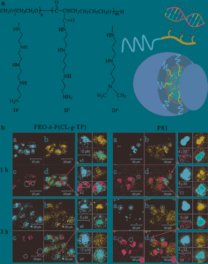
Fig. 17
(
a
) Structures of PEO-P(CL-polyamine) copolymers and siRNA complexed PEO-
P(CL-polyamine) micelles. (
b
) Assessment of endosomal escape for PEO-
b
-P(CL-
g
-TP) and PEI
after intracellular uptake by endocytosis upon 1 and 3 h incubation by confocal microscopy. The
cells were treated with FAM-labeled siRNA formulated in PEO-
b
-P(CL-
g
-TP) and PEI (
green,
panel a
); Lysosomes and nuclei were stained by LysoTracker (
red, panel b
) and DAPI (
blue, panel c
),
respectively, and the images were merged in
panel d
. The endosomes/lysosomes in cells treated
with PCI micelles and particles in different phases were magnified in e and f, respectively
(Reproduced from Xiong et al.
2008b
with permission)
Search WWH ::

Custom Search